. 2016 Dec;35(4):319-28.
doi: 10.3109/15569527.2015.1127255. Epub 2016 Mar 22.

Clinical progression of ocular injury following arsenical vesicant lewisite exposure

Affiliations

Clinical progression of ocular injury following arsenical vesicant lewisite exposure

Neera Tewari-Singh et al. Cutan Ocul Toxicol. 2016 Dec.

Abstract

Ocular injury by lewisite (LEW), a potential chemical warfare and terrorist agent, results in edema of eyelids, inflammation, massive corneal necrosis and blindness. To enable screening of effective therapeutics to treat ocular injury from LEW, useful clinically-relevant endpoints are essential. Hence, we designed an efficient exposure system capable of exposing up to six New-Zealand white rabbits at one time, and assessed LEW vapor-induced progression of clinical ocular lesions mainly in the cornea. The right eye of each rabbit was exposed to LEW (0.2 mg/L) vapor for 2.5, 5.0, 7.5 and 10.0 min and clinical progression of injury was observed for 28 days post-exposure (dose-response study), or exposed to same LEW dose for 2.5 and 7.5 min and clinical progression of injury was observed for up to 56 days post-exposure (time-response study); left eye served as an unexposed control. Increasing LEW exposure caused corneal opacity within 6 h post-exposure, which increased up to 3 days, slightly reduced thereafter till 3 weeks, and again increased thereafter. LEW-induced corneal ulceration peaked at 1 day post-exposure and its increase thereafter was observed in phases. LEW exposure induced neovascularization starting at 7 days which peaked at 22-35 days post-exposure, and remained persistent thereafter. In addition, LEW exposure caused corneal thickness, iris redness, and redness and swelling of the conjunctiva. Together, these findings provide clinical sequelae of ocular injury following LEW exposure and for the first time establish clinically-relevant quantitative endpoints, to enable the further identification of histopathological and molecular events involved in LEW-induced ocular injury.

Keywords: Iris redness; Lewisite; clinical lesions; conjunctival injury; corneal opacity; corneal thickness; corneal ulceration; efficient exposure system; neovascularization.


Figures

Figure 1
Figure 1. LEW vapor system for ocular exposure with the capacity to expose up to six rabbits simultaneously
Exposure system setup under the safety cabinet (A), and LEW ocular exposure of rabbits (B).
Figure 2
Figure 2. Clinical progression of ocular injury following LEW exposure
New Zealand white rabbits (n=5/group) were exposed to LEW (0.2 mg/L) vapor for either 10 min and clinical progression of injury was observed for 7 days post-exposure, or for 2.5 and 5.0 min and clinical progression of injury was observed for up to 56 days post-exposure as detailed in materials and methods. The right eye was exposed to LEW vapor and at the same time, the left eye was exposed to dilution air (no LEW) alone. Representative pictures show the injury to rabbit eyes from the highest (10 min) LEW exposure time at 1 and 7 days post-exposure (A) and the progression of ocular injury from 1 day to 56 days following 2.5 (B) and 7.5 (C) min LEW exposure. Red arrows, Inflammation: swelling and edema in the eyelid and conjunctiva and iris redness; blue arrows, corneal-stromal injury (opacity); purple arrows, neovascularization
Figure 3
Figure 3. Effect of LEW vapor on corneal opacity
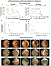
New Zealand white rabbits were exposed to LEW (0.2 mg/L) vapor for either 2.5, 5.0, 7.5 and 10 min and corneal opacity was evaluated from 6 h (Day 0) to 28 days post-exposure (A), or for 2.5 and 7.5 min and corneal opacity was evaluated from 1 day to 56 days post-exposure (B) as detailed in materials and methods. Representative slit-lamp pictures show corneal opacity in rabbit eyes from 1 day to 56 days following 2.5 and 7.5 min LEW exposure (C). Blue arrows, scarring or clouding of cornea or corneal opacity. Data presented are mean ± SEM (n=3-10). *, p<0.05 for 2.5 min LEW exposure; †, p<0.05 for 5.0 min LEW exposure; ‡, p<0.05 for 7.5 min LEW exposure; §, p<0.05 for 10.0 min LEW exposure.
Figure 4
Figure 4. Effect of LEW vapor on corneal ulceration
New Zealand white rabbits were exposed to LEW (0.2 mg/L) vapor for either 2.5, 5.0, 7.5 and 10 min and corneal ulceration was evaluated from 6 h (Day 0) to 28 days post-exposure (A), or for 2.5 and 7.5 min and corneal ulceration was evaluated from 1 day to 56 days post-exposure (B) as detailed in materials and methods. Representative slit-lamp pictures show corneal ulceration in rabbit eyes from 1 day to 56 days following 2.5 and 7.5 min LEW exposure (C). Blue arrows, corneal ulceration; fluorescein stain taken up by exposed stroma, due to the disruption of epithelial layer, appears green. Data presented are mean ± SEM (n=3-10). *, p<0.05 for 2.5 min LEW exposure; †, p<0.05 for 5.0 min LEW exposure; ‡, p<0.05 for 7.5 min LEW exposure; §, p<0.05 for 10.0 min LEW exposure.
Figure 5
Figure 5. Effect of LEW vapor on corneal neovascularization
New Zealand white rabbits were exposed to LEW (0.2 mg/L) vapor for either 2.5, 5.0, 7.5 and 10 min and corneal neovascularization was evaluated from 6 h (Day 0) to 28 days post-exposure (A), or for 2.5 and 7.5 min and corneal neovascularization was evaluated from 1 day to 56 days post-exposure (B) as detailed in materials and methods. Representative slit-lamp pictures show corneal neovascularization in rabbit eyes from 1 day to 56 days following 2.5 and 7.5 min LEW exposure (C). Blue arrows, neovascularization seen as excessive growth of blood vessels. Data presented are mean ± SEM (n=3-10). *, p<0.05 for 2.5 min LEW exposure; †, p<0.05 for 5.0 min LEW exposure; ‡, p<0.05 for 7.5 min LEW exposure; §, p<0.05 for 10.0 min LEW exposure.
Figure 6
Figure 6. Effect of LEW vapor on corneal thickness
New Zealand white rabbits were exposed to LEW (0.2 mg/L) vapor for 2.5 and 7.5 min and corneal thickness was measured using a pachymeter from 1 day to 56 days post-exposure as detailed in materials and methods. Data presented are mean ± SEM (n=3-10). *, p<0.05 for 2.5 min LEW exposure; ‡, p<0.05 for 7.5 min LEW exposure.
Figure 7
Figure 7. Effect of LEW vapor on the conjunctiva
New Zealand white rabbits were exposed to LEW (0.2 mg/L) vapor for either 2.5, 5.0, 7.5 and 10 min and percent animals with conjunctival swelling (A) and redness (C) were evaluated from 6 h (Day 0) to 28 days post-exposure or for 2.5 and 7.5 min and percent animals with conjunctival swelling (B) and redness (D) were evaluated from 1 day to 56 days post-exposure (B) as detailed in materials and methods. Representative slit-lamp pictures show redness and swelling in the conjunctiva of rabbit eyes from 1 day to 56 days following 2.5 and 7.5 min LEW exposure (n=3-10, E). Blue arrows, redness and swelling in the conjunctiva.
Figure 8
Figure 8. Effect of LEW vapor on the iris
New Zealand white rabbits were exposed to LEW (0.2 mg/L) vapor for either 2.5, 5.0, 7.5 and 10 min and percent animals with injury to the iris were evaluated from 6 h (Day 0) to 28 days post-exposure (A), or for 2.5 and 7.5 min and percent animals with injury to the iris were evaluated from 1 day to 56 days post-exposure (B) as detailed in materials and methods. Representative slit-lamp pictures show redness in the iris of rabbit eyes from 1 day to 56 days following 2.5 and 7.5 min LEW exposure (n=3-10, C). Blue arrows, redness and swelling in the conjunctiva.

References

    1. Lindsay CD, Hambrook JL, Brown RF, Platt JC, Knight R, Rice P. Examination of changes in connective tissue macromolecular components of large white pig skin following application of Lewisite vapour. Journal of applied toxicology : JAT. 2004;24(1):37–46. - PubMed
    1. Sahu C, Pakhira S, Sen K, Das AK. A computational study of detoxification of lewisite warfare agents by British anti-lewisite: catalytic effects of water and ammonia on reaction mechanism and kinetics. The journal of physical chemistry A. 2013;117(16):3496–506. Epub 2013/04/02. - PubMed
    1. Kehe K, Flohe S, Krebs G, Kreppel H, Reichl FX, Liebl B, et al. Effects of Lewisite on cell membrane integrity and energy metabolism in human keratinocytes and SCL II cells. Toxicology. 2001;163(2-3):137–44. Epub 2001/08/23. - PubMed
    1. King JR, Riviere JE, Monteiro-Riviere NA. Characterization of lewisite toxicity in isolated perfused skin. Toxicology and applied pharmacology. 1992;116(2):189–201. Epub 1992/10/01. - PubMed
    1. Nelson P, Hancock JR, Sawyer TW. Therapeutic effects of hypothermia on Lewisite toxicity. Toxicology. 2006;222(1-2):8–16. Epub 2006/02/21. - PubMed

MeSH terms