Review
. 2016 Jun;1374(1):17-28.
doi: 10.1111/nyas.13028. Epub 2016 Mar 22.

Long-term neuropathological and behavioral impairments after exposure to nerve agents

Affiliations
Review

Long-term neuropathological and behavioral impairments after exposure to nerve agents

Vassiliki Aroniadou-Anderjaska et al. Ann N Y Acad Sci. 2016 Jun.

Abstract

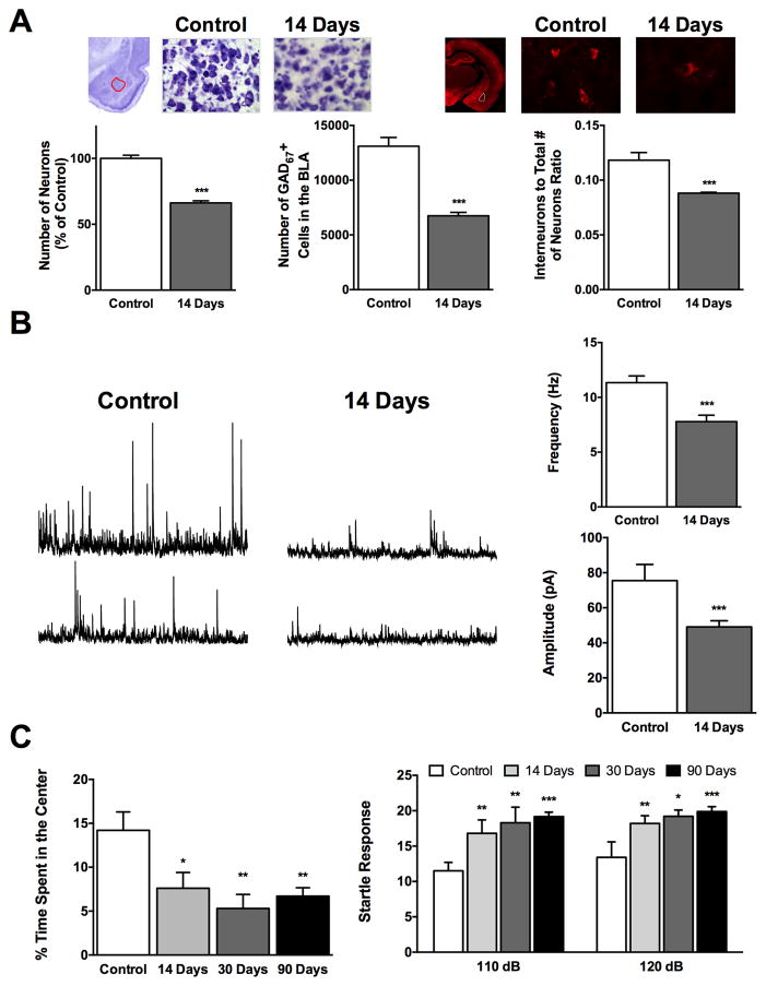
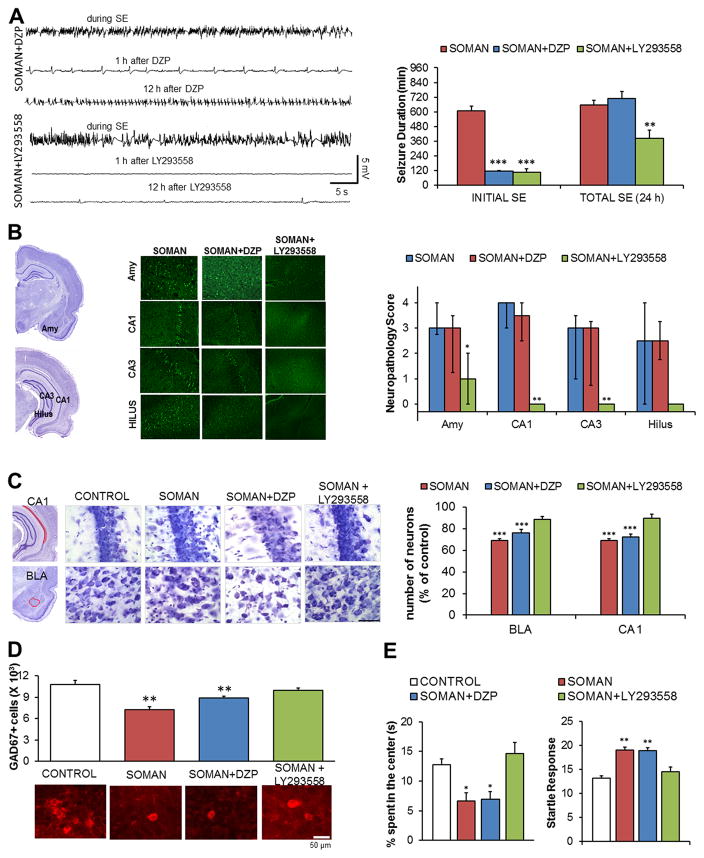
One of the deleterious effects of acute nerve agent exposure is the induction of status epilepticus (SE). If SE is not controlled effectively, it causes extensive brain damage. Here, we review the neuropathology observed after nerve agent-induced SE, as well as the ensuing pathophysiological, neurological, and behavioral alterations, with an emphasis on their time course and longevity. Limbic structures are particularly vulnerable to damage by nerve agent exposure. The basolateral amygdala (BLA), which appears to be a key site for seizure initiation upon exposure, suffers severe neuronal loss; however, GABAergic BLA interneurons display a delayed death, perhaps providing a window of opportunity for rescuing intervention. The end result is a long-term reduction of GABAergic activity in the BLA, with a concomitant increase in spontaneous excitatory activity; such pathophysiological alterations are not observed in the CA1 hippocampal area, despite the extensive neuronal loss. Hyperexcitability in the BLA may be at least in part responsible for the development of recurrent seizures and increased anxiety, while hippocampal damage may underlie the long-term memory impairments. Effective control of SE after nerve agent exposure, such that brain damage is also minimized, is paramount for preventing lasting neurological and behavioral deficits.

Keywords: anxiety; basolateral amygdala; hippocampus; nerve agents; seizures; status epilepticus.


Conflict of interest statement

Conflicts of interest

The authors declare no conflicts of interest.

Figures

Figure 1
Figure 1
Status epilepticus (SE) induced by exposure to soman causes severe neuronal loss in the basolateral amygdala (BLA) that leads to long-term increases in the excitability of the BLA network and increased anxiety-like behavior. (A) Fourteen days after exposure of rats to soman (1.2 × LD50), there is a significant reduction in the total number of neurons—as determined by unbiased design-based stereological counting in Nissl-stained sections—and GABAergic interneurons in the BLA, with a decrease in the ratio of GABAergic to the total number of neurons in comparison to control rats. Photomicrographs in the top show Nissl-stained (left) and GAD-67 labeled (right) sections from the BLA of control rats and soman-exposed rats, 14 days after the exposure. Bar graphs show the group data on neuronal loss (data are from Ref. 24). (B) The frequency and amplitude of spontaneous IPSCs are significantly reduced 14 days after soman-induced SE; representative traces are shown on the left and group data on the right (from Ref. 41). (C) Anxiety-like behavior is increased after soman exposure, an impairment that persists at 90 days postexposure. The time spent in the center of the open field is shown on the left, and the amplitude of the startle responses to acoustic stimuli on the right (from Refs. and 24).
Figure 2
Figure 2
Effective control of SE is necessary for the prevention of neuropathology and behavioral deficits. The data presented here compare the efficacies of LY293558 (an AMPA/GluK1 receptor antagonist) and diazepam (DZP) against soman. (A) Both LY293558 and DZP, administered 1 h after exposure of rats to soman (1.4 × LD50), terminated the initial SE, but seizures returned in the DZP-treated rats; as a result, only LY293558 reduced the total duration of SE within 24 h after the exposure. Electroencephalographic traces are shown on the left and group data on the right (from Refs. and 15). (B) Neuronal degeneration is prevented by LY293558, but not by DZP treatment. Data shown were obtained 7 days after the exposure. The panoramic Nissl-stained sections (left) show the areas where neurodegeneration was evaluated, representative photomicrographs of Fluoro–Jade C–stained sections from the amygdala (Amy) and the hippocampus are shown in the middle, and group data are shown on the right (from Refs. and 15). (C) Neuronal loss in the BLA and the CA1 hippocampal area is prevented by LY293558, but not by DZP treatment. Data were obtained 7 days after the exposure. The panoramic Nissl-stained sections (left) show the areas where neuronal loss was assessed. The Nissl-stained sections in the middle (total magnification 630×; scale bar, 50 μm) are representative photomicrographs from the four groups. The bar graph (right) shows group data (from Refs. and 15). (D) Loss of GABAergic interneurons in the BLA is prevented by LY293558, but not by DZP treatment. Data were obtained 7 days after the exposure. The bar graph shows group data; representative photomicrographs from the corresponding groups are shown below the graph (from Refs. and 15).(E) Increased anxiety-like behavior, 30 days after soman exposure, was prevented by LY293558, but not by DZP treatment. Left graph: time spent in the center of the open field. Right graph: amplitude of the startle responses to 120-dB acoustic stimulus (from Refs. and 15).

References

    1. Sirin GS, Zhou Y, Lior-Hoffmann L, et al. Aging mechanism of soman inhibited acetylcholinesterase. J Phys Chem B. 2012;116:12199–12207. - PMC - PubMed
    1. Bajgar J, Fusek J, Kassa J, et al. An attempt to assess functionally minimal acetylcholinesterase activity necessary for survival of rats intoxicated with nerve agents. Chem Biol Interact. 2008;175:281–285. - PubMed
    1. Shih TM, Kan RK, McDonough JH. In vivo cholinesterase inhibitory specificity of organophosphorus nerve agents. Chem Biol Interact. 2005;157–158:293–303. - PubMed
    1. Fountain NB. Status epilepticus: risk factors and complications. Epilepsia. 2000;41(Suppl 2):S23–30. - PubMed
    1. Schmued LC, Stowers CC, Scallet AC, et al. Fluoro-Jade C results in ultra high resolution and contrast labeling of degenerating neurons. Brain Res. 2005;1035:24–31. - PubMed

Publication types