. 2016 Mar 22;11(3):e0152124.
doi: 10.1371/journal.pone.0152124. eCollection 2016.

Early Impairment of Lung Mechanics in a Murine Model of Marfan Syndrome

Affiliations

Early Impairment of Lung Mechanics in a Murine Model of Marfan Syndrome

Juan J Uriarte et al. PLoS One. .

Abstract

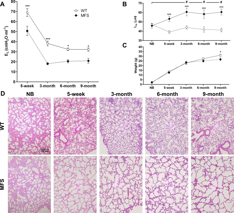
Early morbidity and mortality in patients with Marfan syndrome (MFS) -a connective tissue disease caused by mutations in fibrillin-1 gene- are mainly caused by aorta aneurysm and rupture. However, the increase in the life expectancy of MFS patients recently achieved by reparatory surgery promotes clinical manifestations in other organs. Although some studies have reported respiratory alterations in MFS, our knowledge of how this connective tissue disease modifies lung mechanics is scarce. Hence, we assessed whether the stiffness of the whole lung and of its extracellular matrix (ECM) is affected in a well-characterized MFS mouse model (FBN1C1039G/+). The stiffness of the whole lung and of its ECM were measured by conventional mechanical ventilation and atomic force microscopy, respectively. We studied 5-week and 9-month old mice, whose ages are representative of early and late stages of the disease. At both ages, the lungs of MFS mice were significantly more compliant than in wild type (WT) mice. By contrast, no significant differences were found in local lung ECM stiffness. Moreover, histopathological lung evaluation showed a clear emphysematous-like pattern in MFS mice since alveolar space enlargement was significantly increased compared with WT mice. These data suggest that the mechanism explaining the increased lung compliance in MFS is not a direct consequence of reduced ECM stiffness, but an emphysema-like alteration in the 3D structural organization of the lung. Since lung alterations in MFS are almost fully manifested at an early age, it is suggested that respiratory monitoring could provide early biomarkers for diagnosis and/or follow-up of patients with the Marfan syndrome.


Conflict of interest statement

Competing Interests: The authors have declared that no competing interests exist. Please note that "CIBER Enfermedades Respiratorias" is not a commercial company. It is a públic network of Research in Spain (intrated in the Health Ministry). The whole name in English is “Network Research Center of Respiratory Diseases”. No author is employed by a commercial company.

Figures

Fig 1
Fig 1. Mechanics in 9-month old mice.
(A) Static (Est) and (B) dynamic (Edyn) elastances in WT (white column) and Marfan mice (grey column) determined by the end-inspiratory airway occlusion method. *: p < 0.05 when comparing WT and Marfan. (C) Young’s modulus (E) measured at different regions of the lung ECM in WT (white column) and Marfan mice (grey column). #: p < 0.05, and •: p < 0.05 when comparing different regions in WT and Marfan, respectively. (D) Complex shear modulus (G*) measured at the alveolar septum of decellularized lung matrix in WT (open symbols) and MFS (closed symbols) mice. Circles represent the real part (storage modulus, G’) and triangles represent the imaginary part (loss modulus, G”) of G*. Solid and dashed lines represent fits of the two power law model to G’ and G”, respectively (WT black and MFS grey). Data are mean ± SE.
Fig 2
Fig 2. Mechanics in 5-week old mice.
(A) Static (Est) and (B) dynamic (Edyn) elastances in WT (white column) and Marfan mice (grey column) determined by the end-inspiratory airway occlusion method. (C) Young’s modulus (E) measured at different regions of the lung ECM in WT (white column) and Marfan mice (grey column). (D) Complex shear modulus (G*) measured at the alveolar septum of decellularized lung matrix in WT (open symbols) and MFS (closed symbols) mice. Circles represent the real part (storage modulus, G’) and triangles represent the imaginary part (loss modulus, G”) of G*. Solid and dashed lines represent fits of the two power law model to G’ and G”, respectively (WT black and MFS grey). Data are mean ± SE. *: p < 0.05.
Fig 3
Fig 3. Lung elastances before and after decellularization.
(A) Static (Est) and (B) dynamic (Edyn) elastances in WT (white column) and Marfan mice (MFS; grey column) determined by the end-inspiratory airway occlusion method in vivo and in the acellular lung scaffolds after organ decellularization. Data are mean ± SE. *: p < 0.05; **: p < 0.01.
Fig 4
Fig 4. Histological analysis of mouse lungs.
Representative H&E stained images of lung sections from (A) 9-month and (B) 5-week old wild-type (WT), and (C) 9-month and (D) 5-week old Marfan (MFS) mice. Images acquired at 100x magnification and scale bar represents 200μm. (E) Mean linear intercept (Lm) of alveolar septa. Data are mean ± SE. * p < 0.05; *** p <0.001 compared with their respective WT and # p < 0.05 between different ages in Marfan mice.
Fig 5
Fig 5. Airspace enlargement evolution in WT and MFS mice.
(A) Effective lung elastance (EL) and (B) mean linear interecept (Lm) of alveolar septa in mice of different ages: from newborn (NB) up to 9-month old in wild-type (WT; open circles) and Marfan (MFS; closed black circles) mice. (C) Body weight along age in MFS and WT mice. (D) Panel illustrates examples of H&E stained lung sections. Images acquired at 100x magnification and scale bar represents 200μm. Data are mean ± SE. ***: p<0.001 between WT and Marfan animals and #: p<0.05 respective to new born mice in Marfan animals.
Fig 6
Fig 6. Tunica media fibers integrity in WT and MFS mice aortas.
(A) Representative images from ascending aortas in WT and Marfan (MFS) mice processed and evaluated for elastic tunica media fibers integrity. Histological sections were stained with Verhoeff-Van Gieson. Images acquired at 40x magnification and scale bar represents 50μm.(B) Elastic fiber ruptures in the tunica media of the ascending aorta. Data are mean ± SE. *: p<0.05 between WT and Marfan animals

References

    1. von Kodolitsch Y, De Backer J, Schüler H, Bannas P, Behzadi C, Bernhardt AM, et al. Perspectives on the revised Ghent criteria for the diagnosis of Marfan syndrome. Appl Clin Genet. 2015;8: 137–55. 10.2147/TACG.S60472 - DOI - PMC - PubMed
    1. Cañadas V, Vilacosta I, Bruna I, Fuster V. Marfan syndrome. Part 1: pathophysiology and diagnosis. Nat Rev Cardiol. 2010;7: 256–65. 10.1038/nrcardio.2010.30 - DOI - PubMed
    1. Dietz HC, Cutting GR, Pyeritz RE, Maslen CL, Sakai LY, Corson GM,et al. Marfan syndrome caused by a recurrent de novo missense mutation in the fibrillin gene. Nature. 1991;352: 337–39. - PubMed
    1. Lima BL, Santos EJ, Fernandes GR, Merkel C, Mello MR, Gomes JPA,et al. A new mouse model for marfan syndrome presents phenotypic variability associated with the genetic background and overall levels of Fbn1 expression. PLoS One 2010;5: e14136 10.1371/journal.pone.0014136 - DOI - PMC - PubMed
    1. Pereira L, Andrikopoulos K, Tian J, Lee SY, Keene DR, Ono R, et al. Targetting of the gene encoding fibrillin-1 recapitulates the vascular aspect of Marfan syndrome. Nat Genet 1997;17: 218–22. - PubMed

Publication types