Review
. 2016 Mar;11(6):673-92.
doi: 10.2217/nnm.16.5. Epub 2016 Mar 22.

The effect of nanoparticle size on in vivo pharmacokinetics and cellular interaction

Affiliations
Review

The effect of nanoparticle size on in vivo pharmacokinetics and cellular interaction

Nazanin Hoshyar et al. Nanomedicine (Lond). 2016 Mar.

Abstract

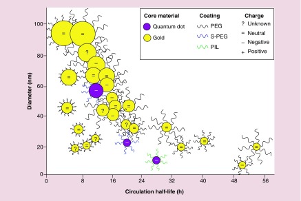
Nanoparticle-based technologies offer exciting new approaches to disease diagnostics and therapeutics. To take advantage of unique properties of nanoscale materials and structures, the size, shape and/or surface chemistry of nanoparticles need to be optimized, allowing their functionalities to be tailored for different biomedical applications. Here we review the effects of nanoparticle size on cellular interaction and in vivo pharmacokinetics, including cellular uptake, biodistribution and circulation half-life of nanoparticles. Important features of nanoparticle probes for molecular imaging and modeling of nanoparticle size effects are also discussed.

Keywords: cellular uptake; in vivo pharmacokinetics; modeling; nanoparticle; size effect.


Conflict of interest statement

Financial & competing interests disclosure

This work was supported by the National Heart Lung and Blood Institute of the NIH as a Program of Excellence in Nanotechnology award (HHSN268201000043C to GB). The authors have no other relevant affiliations or financial involvement with any organization or entity with a financial interest in or financial conflict with the subject matter or materials discussed in the manuscript apart from those disclosed.

No writing assistance was utilized in the production of this manuscript.

Figures

<b>Figure 1.</b>
Figure 1.. A schematic depicting the effect of nanoparticle size on the membrane-wrapping process.
(A) Nanoparticles greater than 60 nm in diameter driving the membrane-wrapping process by binding to a large number of receptors but limiting the binding of other nanoparticles. (B) Nanoparticles less than 30 nm in diameter attaching to some membrane receptors but failing to drive the membrane-wrapping process unless many bind to receptors in close proximity. (C) Nanoparticles between 30 and 60 nm in diameter attaching to membrane receptors and driving the membrane-wrapping process effectively.
<b>Figure 2.</b>
Figure 2.. A schematic representing the relationship between nanoparticle size and circulation half-life.
Each point represents a specific combination of characteristics that has been tested. Points are colored based on core material (yellow represents gold and purple represents quantum dots). The coating of the particle is shown by the lines surrounding each point. Black lines denote a PEG coating, with longer lines representing higher PEG molecular weight. Blue lines denote an S-PEG coating (silica shell with PEG5000 coating). Green lines denote a PIL coating. Within each point, surface charge is shown as positive (+), negative (-), neutral (=), or unknown (?).
<b>Figure 3.</b>
Figure 3.. Biodistribution of nanoparticles showing the effect of size.
(A) Biodistribution of 13N-labeled Al2O3 nanoparticles in male rats 60 min postinjection. Graphs show percentage ID that has accumulated in the brain (top left), liver (bottom left), lungs (top right) and kidneys (bottom right). (B) Biodistribution of bare and TPGS-coated fluorescent PS NPs in Sprague–Dawley rats 3 h postinjection. ID: Injected dose; PS: Polystyrene nanoparticle. (A) Reproduced with permission from [60]. (B) Reproduced with permission from [5].
<b>Figure 4.</b>
Figure 4.. Size-dependent relaxivities of nanoparticle probes.
(A) r1 relaxivity for magnetite in various oxidation states. Each shows a positive, linear correlation between nanoparticle size and r1 relaxivity. (B) T2 relaxivity of the SPIONs on a per particle basis. SPIONs with two core sizes, 5 and 14 nm, and five PEG sizes, molecular weight of 550, 750, 1000, 2000 and 5000 Da, were evaluated. (C) MRI images before (pre) and after (post) magnetic nanoparticle injection into the foot on the hind leg of mice with a subcutaneous SCCVII tumor. A 3T scanner was used. (A) Reprinted with permission from [14]. (B) Reproduced with permission from [13]. (C) Reproduced with permission from [83].
<b>Figure 5.</b>
Figure 5.. Schematics of physiologically based pharmacokinetic models for nanoparticle permeability.
(A) A perfusion model (left) and permeability model (right) with the solid triangle indicating intravenous injection, arrows indicating nanoparticle transportation direction and dashed arrows indicating transportation equations in the permeability model that differ from those in the perfusion model. (B) A schematic of a more complex permeability model that considers the effects of phagocytosis on nanoparticle distribution. iv.: Intravenous. (A) Reproduced with permission from [70]. (B) Reproduced with permission from [90].

References

    1. Albanese A, Tang PS, Chan WCW. The effect of nanoparticle size, shape, and surface chemistry on biological systems. Ann. Rev. Biomed. Eng. 2012;14:1–16. - PubMed
    1. Mitragotri S, Anderson DG, Chen X, et al. Accelerating the translation of nanomaterials in biomedicine. ACS Nano. 2015;9:6644–6654. - PMC - PubMed
    1. de Barros AB, Tsourkas A, Saboury B, Cardoso VN, Alavi A. Emerging role of radiolabeled nanoparticles as an effective diagnostic technique. EJNMMI Res. 2012;2:39–53. - PMC - PubMed
    1. Elias DR, Poloukhtine A, Popik V, Tsourkas A. Effect of ligand density, receptor density, and nanoparticle size on cell targeting. Nanomedicine. 2013;9:194–201. - PMC - PubMed
    1. Kulkarni SA, Feng SS. Effects of particle size and surface modification on cellular uptake and biodistribution of polymeric nanoparticles for drug delivery. Pharmaceut. Res. 2013;30:2512–2522. - PubMed

Publication types