Review
. 2016 Mar 21;26(6):R247-56.
doi: 10.1016/j.cub.2016.02.034.

The Eukaryotic Replisome Goes Under the Microscope

Affiliations
Review

The Eukaryotic Replisome Goes Under the Microscope

Mike O'Donnell et al. Curr Biol. .

Abstract

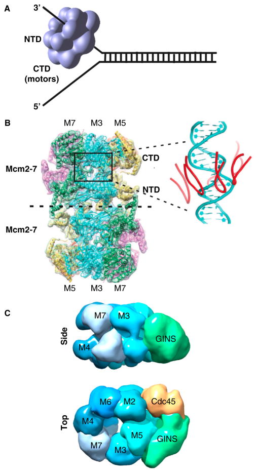
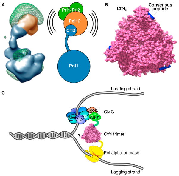
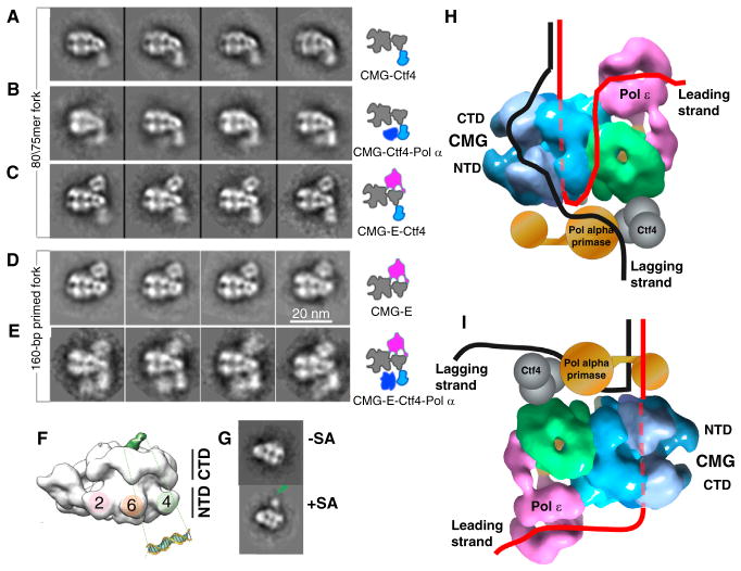
The machinery at the eukaryotic replication fork has seen many new structural advances using electron microscopy and crystallography. Recent structures of eukaryotic replisome components include the Mcm2-7 complex, the CMG helicase, DNA polymerases, a Ctf4 trimer hub and the first look at a core replisome of 20 different proteins containing the helicase, primase, leading polymerase and a lagging strand polymerase. The eukaryotic core replisome shows an unanticipated architecture, with one polymerase sitting above the helicase and the other below. Additionally, structures of Mcm2 bound to an H3/H4 tetramer suggest a direct role of the replisome in handling nucleosomes, which are important to DNA organization and gene regulation. This review provides a summary of some of the many recent advances in the structure of the eukaryotic replisome.


Figures

Figure 1
Figure 1. The eukaryotic CMG helicase
(A) Steric exclusion model of helicase unwinding. All replicative helicases contain hexameric motor rings that can encircle and translocate along one strand of ssDNA, excluding the other strand. Individual subunits of hexameric helicases are composed of amino- and carboxy-terminal domains (NTD and CTD, respectively); the ATP site is in the CTD. (B) CryoEM atomic structure of the Mcm2-7 double hexamer of S. cerevisiae. The inset, right, shows the six h2i loops in the interior channel that closely approach DNA modeled into the structure. Reprinted by permission from Macmillan Publishers Ltd: Nature [40], copyright 2015. (C) EM 3D reconstruction of S. cerevisiae CMG in side and carboxy-terminal top views. Representations were of EMD 6463 from [43] using Chimera.
Figure 2
Figure 2. Pol alpha–primase and the Ctf4 trimer
(A) EM 3D reconstruction of the four-subunit Pol alpha from S. cerevisiae. The Pol1 polymerase subunit contains a CTD linked by a flexible tether to which the other subunits attach. Adapted from [59] by permission of Oxford University Press. (B) Surface representation of the C-half of S. cerevisiae Ctf4 trimer (magenta), with bound peptides modeled into the peptide binding pockets (blue). Drawn using Chimera and PDB 4CBH from [61]. (C) Cartoon of a Ctf4 trimer binding GINS within CMG (leading strand), and Pol alpha, inferred from Ctf4–peptide interactions. One subunit of Ctf4 may interact with other binding partners yet to be identified (question mark). Both B and C are reprinted by permission from Macmillan Publishers Ltd: Nature [61], copyright 2014.
Figure 3
Figure 3. The leading strand CMGE replisome
(A) EM 2D averages of S. cerevisiae CMG are in the top row and CMGE (i.e. CMG + Pol epsilon) is in the bottom row. (B) 3D reconstruction of S. cerevisiae CMGE. CMGE is drawn with Chimera using EMD-6465 from [43].
Figure 4
Figure 4. Core replisome architecture
(A–E) 2D class averages of complexes as depicted in the illustrations to the right of each row of images. Adapted from Figure 5 of [42]. (F) DNA path through CMG determined in the Drosophila system [79]. The DNA duplex region (green) was visible in the negative stain 3D reconstruction. (G) This was confirmed by a streptavidin-biotin tag (SA) on the 20 bp duplex. Both F and G are adapted with permission from Figure 2 of [79]. (H) Combining the DNA path from the Drosophila CMG–DNA structure (79) with the S. cerevisiae CMGE structure and the class averages of panels A–E [42] places the Pol epsilon on the top (C-face) of CMG adjacent to the parental duplex. After the unwound leading strand exits the NTD of CMG, it needs to make a U-turn to reach Pol epsilon on the top, illustrated as taking an external path, but the ssDNA may take an internal path through CMG. In the steric exclusion model, the lagging strand is on the outside of CMG to reach Pol alpha on the N-side. CMGE was drawn with Chimera using EMD-6465 from [42]. (I) If DNA were to enter the NTD of the Mcm2-7, the Pol epsilon would reside below the CMG and Pol alpha primase would ride near the DNA split point, where it could prime the lagging strand.
Figure 5
Figure 5. Mcm2 binds the H3/H4 heterotetramer
(A) Crystal structure of the amino-terminal histone binding region of Mcm2 (pink) with the H3/H4 tetramer (green and blue) drawn using Chimera and PDB 5BNV from [85]. (B) Crystal structure of the nucleosome drawn using Chimera and PDB 1A0I from [90]. Histones H3 and H4 are in green and blue. Histones H2A and H2B are in orange and yellow. The location at which Mcm2 binds is not depicted but can be inferred by comparison to panel A. (C) Crystal structure of the Mcm2 histone binding region (pink) with a H3/H4 heterodimer (green and blue) and Asf1 (black) drawn using Chimera and PDB 5BNX from [85]. Figure reprinted by permission from Macmillan Publishers Ltd: Nature Structural and Molecular Biology [85], copyright 2015.

References

    1. Watson JD, Crick FH. Genetical implications of the structure of deoxyribonucleic acid. Nature. 1953;171:964–967. - PubMed
    1. Kornberg A, Baker TA. DNA Replication. New York: W.H. Freeman; 1992.
    1. Barros T, Guenther J, Kelch B, Anaya J, Prabhakar A, O’Donnell M, Kuriyan J, Lamers MH. A structural role for the PHP domain in E. coli DNA polymerase III. BMC Struct Biol. 2013;13:8. - PMC - PubMed
    1. Benkovic SJ, Valentine AM, Salinas F. Replisome-mediated DNA replication. Annu Rev Biochem. 2001;70:181–208. - PubMed
    1. Yao NY, O’Donnell ME. Comparison of bacterial and eukaryotic replisome components. In: Bradshaw RA, Stahl PD, editors. Encyclopedia of Cell Biology. Academic Press; 2015. pp. 396–417.

Publication types

MeSH terms

LinkOut - more resources