

Human tooth and root canal morphology reconstruction using magnetic resonance imaging
- PMID: 27004037
- PMCID: PMC4777457
- DOI: 10.15386/cjmed-555
Human tooth and root canal morphology reconstruction using magnetic resonance imaging
Abstract
Background and aims: Visualization of the internal and external root canal morphology is very important for a successful endodontic treatment; however, it seems to be difficult considering the small size of the tooth and the complexity of the root canal system. Film-based or digital conventional radiographic techniques as well as cone beam computed tomography provide limited information on the dental pulp anatomy or have harmful effects. A new non-invasive diagnosis tool is magnetic resonance imaging, due to its ability of imaging both hard and soft tissues. The aim of this study was to demonstrate magnetic resonance imaging to be a useful tool for imaging the anatomic conditions of the external and internal root canal morphology for endodontic purposes.
Methods: The endodontic system of one freshly extracted wisdom tooth, chosen for its well-known anatomical variations, was mechanically shaped using a hybrid technique. After its preparation, the tooth was immersed into a recipient with saline solution and magnetic resonance imaged immediately. A Bruker Biospec magnetic resonance imaging scanner operated at 7.04 Tesla and based on Avance III radio frequency technology was used. InVesalius software was employed for the 3D reconstruction of the tooth scanned volume.
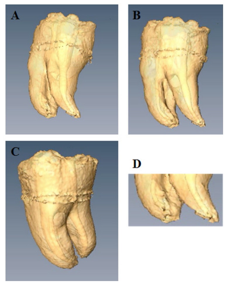
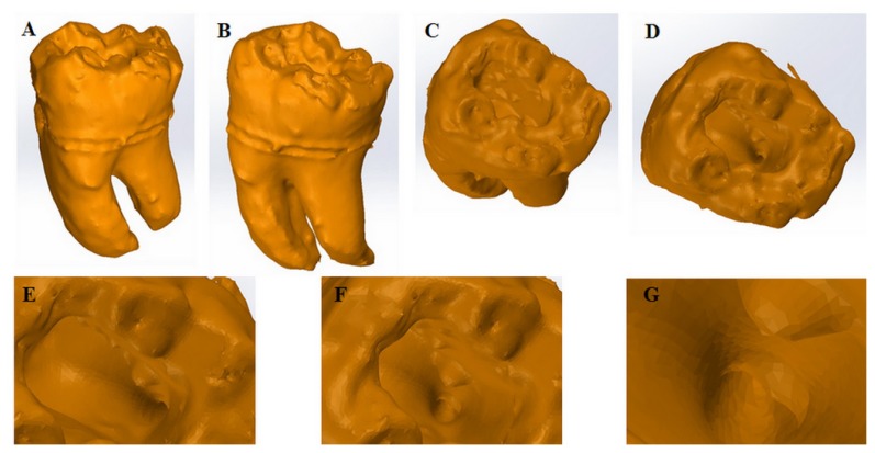
Results: The current ex-vivo experiment shows the accurate 3D volume rendered reconstruction of the internal and external morphology of a human extracted and endodontically treated tooth using a dataset of images acquired by magnetic resonance imaging. The external lingual and vestibular views of the tooth as well as the occlusal view of the pulp chamber, the access cavity, the distal canal opening on the pulp chamber floor, the coronal third of the root canals, the degree of root separation and the apical fusion of the two mesial roots, details of the apical region, root canal curvatures, furcal region and interradicular root grooves could be clearly bordered.
Conclusions: Magnetic resonance imaging offers 3D image datasets with more information than the conventional radiographic techniques. Due to its ability of imaging both hard and soft dental tissues, magnetic resonance imaging can be successfully used as a 3D diagnostic imaging technique in dentistry. When choosing the imaging method, dental clinicians should weight the benefit-risk ratio, taking into account the costs associated to magnetic resonance imaging and the harmful effects of ionizing radiations when cone beam computed tomography or conventional x-ray are used.
Keywords: magnetic resonance imaging; radiographic image enhancement; radiography; root canal; tooth morphology.
Figures
References
-
- Schroeder H. Orale Strukturbiologie. Stuttgart/New York: 1987.
-
- Vertucci FJ. Root canal anatomy of the human permanent teeth. Oral Surg Oral Med Oral Pathol. 1984;58:589–599. - PubMed
-
- Velvart P, Hecker H, Tillinger G. Detection of the apical lesion and the mandibular canal in conventional radiography and computed tomography. Oral Surg Oral Med Oral Pathol Oral Radiol Endod. 2001;92:682–688. - PubMed
-
- Paurazas SB, Geist JR, Pink FE, Hoen MM, Steiman HR. Comparison of diagnostic accuracy of digital imaging by using CCD and CMOS-APS sensors with E-speed film in the detection of periapical bony lesions. Oral Surg Oral Med Oral Pathol Oral Radiol Endod. 2000;89:356–362. - PubMed
-
- Forsberg J, Halse A. Radiographic simulation of a periapical lesion comparing the paralleling and the bisecting-angle techniques. Int Endod J. 1994;27:133–138. - PubMed
LinkOut - more resources
Full Text Sources
Other Literature Sources