

Cadaveric Analysis of the Kambin's Triangle
- PMID: 27004152
- PMCID: PMC4780690
- DOI: 10.7759/cureus.475
Cadaveric Analysis of the Kambin's Triangle
Abstract
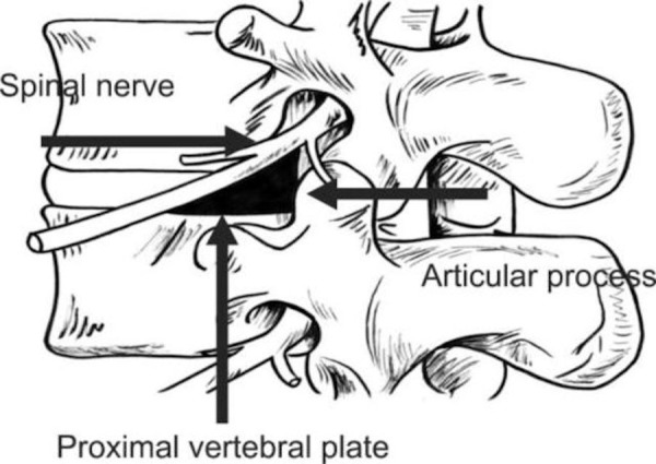
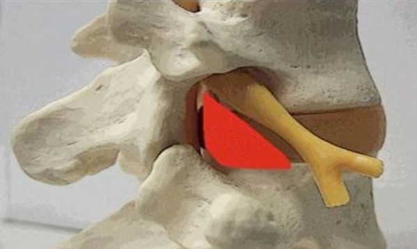
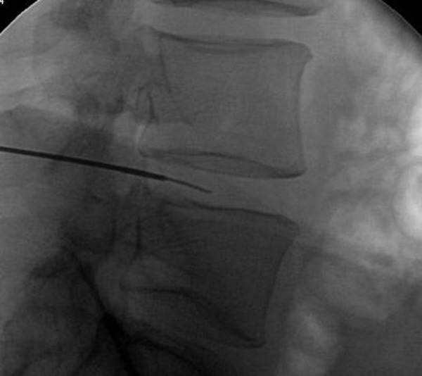
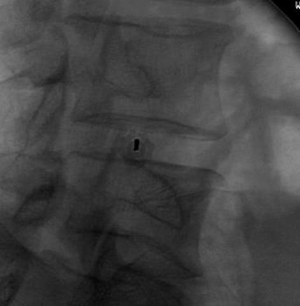
Introduction Kambin's Triangle is a right triangle over the dorsolateral disc. The area of this right triangle currently serves as a strategic site of posterolateral, minimally invasive access to the nerve root for delivery of epidural steroid injections. This posterolateral approach has also been considered a safe area of access to the intervertebral disc space and, thus, an effective approach in reducing complications, such as violation of the thecal sac, the nerve root, or the bony elements of the spine during minimally invasive spinal surgery. No published studies have been performed to characterize the dimensions of the Kambin's Triangle. Our aim is to characterize its dimensions at the lumbar levels and determine its efficacy and safety as a site of access for minimally invasive spinal surgery. Methods Two randomly chosen adult male cadavers were used for this study. The measurements were made bilaterally at their lumbar levels (L1-L5), which equates to 16 total measurements (eight bilateral disc spaces on two cadavers). The disc space was first accessed using a Kirschner wire in a standard oblique approach. With the assistance of fluoroscopy, a Kirschner wire was passed into the disc through the Kambin's Triangle. The procedure was performed on the cadavers bilaterally at four levels, followed by open dissection. The calculations of the area were made by measuring the exiting nerve root, the superior border of the caudal vertebra, and the superior articulating facet-the borders of the Kambin's Triangle. Results The Kambin's Triangle height and width respectively averaged at 12 mm and 10 mm (L1-L2), 13 mm and 11 mm (L2-L3), 17 mm and 11 mm (L3-L4), and 18 mm and 12 mm (L4-L5). Thus, the area at each level was 60 mm(2) (L1-L2), 71.5 mm(2) (L2-L3), 93.5 mm(2) (L3-L4), and 108 mm(2) (L4-L5). All dissected levels demonstrated adequate anchoring of the Kirschner wire into the disc space with no evidence of nerve injury. Following this, a retractor was placed and complete discectomies were performed. All exiting nerves were protected in this safe zone and the thecal sac remained inviolate. Conclusion Understanding the Kambin's Triangle will assist surgeons in the minimally invasive approach to spinal surgeries, with potentially safe placement of interbody cages through this strategic space.
Keywords: kambin; minimally invasive spine surgery; triangle.
Conflict of interest statement
The authors have declared that no competing interests exist.
Figures
References
-
- Posterolateral percutaneous suction-excision of herniated lumbar intervertebral discs: report of interim results. Kambin P, Sampson S. Clinical Orthopaedics and Related Research. 1986;207:37–43. - PubMed
-
- Evaluation of lumbar transforaminal epidural injections with needle placement and contrast flow patterns: a prospective, descriptive report. Manchikanti L, Cash KA, Pampati V, Damron KS, McManus CD. Pain Physician. 2004;7:217–223. - PubMed
-
- Therapeutic spinal corticosteroid injections for the management of radiculopathies. Slipman CW, Chow DW. Physical Medicine and Rehabilitation Clinics of North America. 2002;13:697–711. - PubMed
-
- Fluoroscopically guided lumbar transformational epidural steroid injections in degenerative lumbar stenosis: an outcome study. Botwin KP, Gruber RD, Bouchlas CG, Torres-Ramos FM, Sanelli JT, Freeman ED, Slaten WK, Rao S. American Journal of Physical Medicine & Rehabilitation/Associationof Academic Physiatrists. 2002;81:898–905. - PubMed
LinkOut - more resources
Full Text Sources
Other Literature Sources