. 2016 Sep;27(9):656-67.
doi: 10.1089/hum.2015.160. Epub 2016 May 4.

Long-Term Restoration of Thymidine Phosphorylase Function and Nucleoside Homeostasis Using Hematopoietic Gene Therapy in a Murine Model of Mitochondrial Neurogastrointestinal Encephalomyopathy

Affiliations

Long-Term Restoration of Thymidine Phosphorylase Function and Nucleoside Homeostasis Using Hematopoietic Gene Therapy in a Murine Model of Mitochondrial Neurogastrointestinal Encephalomyopathy

Javier Torres-Torronteras et al. Hum Gene Ther. 2016 Sep.

Abstract

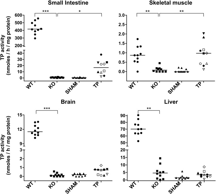
Mitochondrial neurogastrointestinal encephalomyopathy (MNGIE) is a metabolic disorder caused by mutations in TYMP, encoding thymidine phosphorylase (TP). In MNGIE patients, TP dysfunction produces systemic thymidine and deoxyuridine accumulation, which ultimately impairs mitochondrial DNA replication and results in mitochondrial dysfunction. To date, only allogeneic hematopoietic stem cell transplantation has demonstrated long-term clinical efficacy, but high morbidity and mortality associated with this procedure necessitate the search for safer alternatives. In a previous study, we demonstrated that hematopoietic stem cell gene therapy using a lentiviral vector containing the coding sequence of TYMP restored the biochemical homeostasis in an animal model of MNGIE. In the present follow-up study, we show that ectopic expression of TP in the hematopoietic system restores normal nucleoside levels in plasma, as well as in tissues affected in MNGIE such as small intestine, skeletal muscle, brain, and liver. Mitochondrial dNTP pool imbalances observed in liver of the animal model were also corrected by the treatment. The biochemical effects were maintained at least 20 months even with low levels of chimerism. No alterations in the blood cell counts or other toxic effects were observed in association with the lentiviral transduction or TP overexpression. These results further support the notion that gene therapy is a feasible treatment option for MNGIE.


Conflict of interest statement

Author Disclosure No competing financial interests exist.

Figures

<b>Figure 1.</b>
Figure 1.
TP activity in hematopoietic tissues. TP activity in blood, BM, and spleen of wild type (WT), nontreated (KO), p-sham-treated (SHAM), and p-TP-treated (TP) Tymp/Upp1 dKO mice 6 months after treatment. To facilitate matching the results, each mouse in the TP-treated group is identified with the same symbol in Figs. 1–4. Horizontal lines indicate medians. BM, bone marrow; KO, knockout; dKO, double-knockout; TP, thymidine phosphorylase.
<b>Figure 2.</b>
Figure 2.
Molecular chimerism. Molecular chimerism measured through flow cytometry as percentage of green fluorescent cells (a) and lentiviral integrations per cell measured by qRT-PCR, using an EGFP-specific probe referred to the single-copy mouse gene angiogenin-1 (b) in white blood cells, BM, and spleen of sham (n = 10) and TP-treated (n = 10) dKO mice 6 months after treatment. In order to facilitate matching the results, each mouse in the TP-treated group is identified with the same symbol. Horizontal lines indicate median. Statistical analyses were performed with the nonparametric Mann–Whitney U-test (*p < 0.05, **p < 0.01).
<b>Figure 3.</b>
Figure 3.
TP activity in different tissues. TP activity in liver, brain, small intestine, and gastrocnemius muscle from wt (WT), nontreated (KO), p-sham-treated (SHAM), and p-TP-treated (TP) Tymp/Upp1 dKO mice 6 months after treatment. Horizontal lines indicate median. In order to facilitate matching the results, each mouse in the TP-treated group is identified with the same symbol. Statistical analysis were performed with the nonparametric Dunn's multiple comparison test (*p < 0.05, **p < 0.01, ***p < 0.001).
<b>Figure 4.</b>
Figure 4.
Systemic nucleoside clearance. dThd and dUrd concentration in plasma, spleen, brain, liver, skeletal muscle, and small intestine from wt (WT, n = 10), nontreated (KO, n = 10), p-sham-treated (SHAM, n = 10), and p-TP-treated (TP, n = 10) Tymp/Upp1 dKO mice 6 months after treatment. Horizontal lines indicate median. In order to facilitate matching the results, each mouse in the TP-treated group is identified with the same symbol. Statistical analyses were performed with the parametric Dunnett's multiple comparison test (*p < 0.05, **p < 0.01, ***p < 0.001).
<b>Figure 5.</b>
Figure 5.
Survival curve and blood cell counts. (a) Kaplan–Meier survival representation of wt (n = 111), nontreated (n = 181), p-sham-treated (n = 21), p-TP-treated (n = 22), and BM-transplanted (n = 9) dKO mice. (b) Blood cell counts in wt (n = 9), nontreated (n = 10), p-sham-treated (n = 7), and p-TP-treated (n = 5) dKO mice 18 months after treatment (20-month-old mice). Box plots represent the median (horizontal line), interquartile range (box), and minimum and maximum (whiskers), except outliers, which are depicted as dots.
<b>Figure 6.</b>
Figure 6.
Liver mitochondrial dNTP concentrations. Mitochondrial dNTP concentration in liver from wt (WT, n = 10), nontreated (KO, n = 10), p-sham-treated (SHAM, n = 7), and p-TP-treated (TP, n = 5) dKO mice 12 months after treatment. Box plots represent the median (horizontal line), interquartile range (box), and minimum and maximum (whiskers), except outliers, which are depicted as open circles. Asterisks indicate significant differences with WT (*p < 0.05; **p < 0.001; ***p < 0.0001, Student's t-test).

References

    1. Hirano M, Garcia-de-Yebenes J, Jones AC, et al. . Mitochondrial neurogastrointestinal encephalomyopathy syndrome maps to chromosome 22q13.32-qter. Am J Hum Genet 1998;63:526–533 - PMC - PubMed
    1. Hirano M, Nishigaki Y, Marti R. Mitochondrial neurogastrointestinal encephalomyopathy (MNGIE): A disease of two genomes. Neurologist 2004;10:8–17 - PubMed
    1. Hirano M, Silvestri G, Blake DM, et al. . Mitochondrial neurogastrointestinal encephalomyopathy (MNGIE): Clinical, biochemical, and genetic features of an autosomal recessive mitochondrial disorder. Neurology 1994;44:721–727 - PubMed
    1. Desgranges C, Razaka G, Rabaud M, et al. . Catabolism of thymidine in human blood platelets: Purification and properties of thymidine phosphorylase. Biochim Biophys Acta 1981;654:211–218 - PubMed
    1. Marti R, Nishigaki Y, Vila MR, et al. . Alteration of nucleotide metabolism: A new mechanism for mitochondrial disorders. Clin Chem Lab Med 2003;41:845–851 - PubMed

MeSH terms

Supplementary concepts

LinkOut - more resources