

Intrahepatic cholangiocarcinoma in a patient with Wilson's disease: a case report
- PMID: 27005296
- PMCID: PMC4803712
- DOI: 10.1186/s40792-016-0156-3
Intrahepatic cholangiocarcinoma in a patient with Wilson's disease: a case report
Abstract
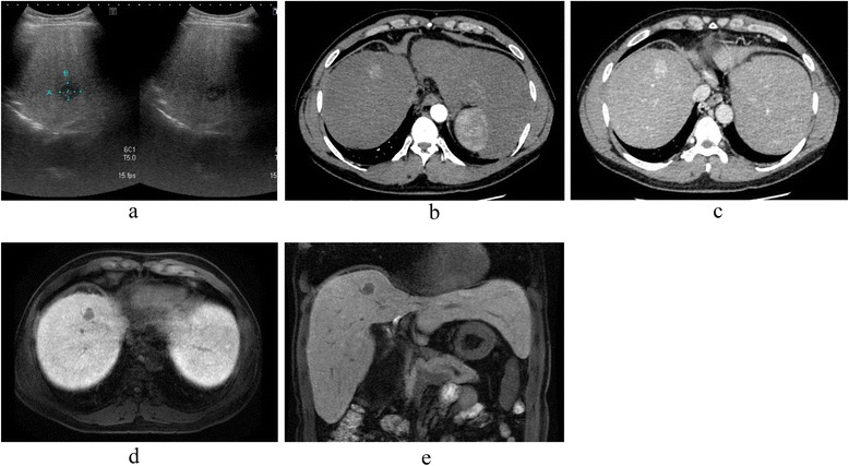
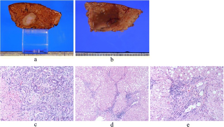
The incidence of hepatobiliary malignancies, and especially intrahepatic cholangiocarcinoma (ICC), for patients with Wilson's disease (WD), is very low, even for cirrhotic patients. A 44-year-old male was admitted to our department for treatment of a liver tumor. He was diagnosed with WD at the age of 15. According to radiological findings, his liver tumor was a suspected hepatocellular carcinoma (HCC) or a combined hepatocellular and cholangiocellular carcinoma. A partial resection of liver segments 8 (S8) and 5 (S5) was subsequently performed due to the intraoperative suspicion of intrahepatic metastasis at the surface of S5. Postoperative histology revealed that the resected portion of S8 contained an ICC; the removed S5 portion comprised a regenerative nodule with hemosiderosis. To date, the patient has survived without tumor recurrence for more than 44 months following surgery. A survey of the literature, inclusive of case reports, would suggest that surgical resection is the primary course of action for a WD patient with ICC, if liver function can be preserved and curative resection performed.
Keywords: Hepatobiliary malignancies; Intrahepatic cholangiocarcinoma; Liver tumor; Wilson’s disease.
Figures
Similar articles
-
Intrahepatic Cholangiocarcinoma in Wilson's Disease: A Case Report.Am J Case Rep. 2024 Jan 27;25:e942372. doi: 10.12659/AJCR.942372. Am J Case Rep. 2024. PMID: 38279525 Free PMC article.
-
A rare case of synchronous double primary hepatocellular carcinoma and intrahepatic cholangiocarcinoma: A case report.Int J Surg Case Rep. 2024 May;118:109599. doi: 10.1016/j.ijscr.2024.109599. Epub 2024 Apr 3. Int J Surg Case Rep. 2024. PMID: 38581941 Free PMC article.
-
Hepatobiliary malignancies in Wilson disease.Liver Int. 2015 May;35(5):1615-22. doi: 10.1111/liv.12727. Epub 2014 Nov 23. Liver Int. 2015. PMID: 25369181
-
Double primary hepatic cancer (hepatocellular carcinoma and intrahepatic cholangiocarcinoma) originating from hepatic progenitor cell: a case report and review of the literature.World J Surg Oncol. 2016 Aug 17;14(1):218. doi: 10.1186/s12957-016-0974-6. World J Surg Oncol. 2016. PMID: 27535234 Free PMC article. Review.
-
Synchronous double cancers of primary hepatocellular carcinoma and intrahepatic cholangiocarcinoma: a case report and review of the literature.World J Surg Oncol. 2014 Nov 10;12:337. doi: 10.1186/1477-7819-12-337. World J Surg Oncol. 2014. PMID: 25385169 Free PMC article. Review.
Cited by
-
Wilson disease, ABCC2 c.3972C > T polymorphism and primary liver cancers: suggestions from a familial cluster.BMC Med Genet. 2020 Nov 18;21(1):225. doi: 10.1186/s12881-020-01165-0. BMC Med Genet. 2020. PMID: 33208122 Free PMC article.
-
Wilson disease and the differential diagnosis of its hepatic manifestations: a narrative review of clinical, laboratory, and liver histological features.Ann Transl Med. 2021 Sep;9(17):1394. doi: 10.21037/atm-21-2264. Ann Transl Med. 2021. PMID: 34733946 Free PMC article. Review.
-
Intrahepatic Cholangiocarcinoma in Wilson's Disease: A Case Report.Am J Case Rep. 2024 Jan 27;25:e942372. doi: 10.12659/AJCR.942372. Am J Case Rep. 2024. PMID: 38279525 Free PMC article.
-
Wilson Disease: Copper-Mediated Cuproptosis, Iron-Related Ferroptosis, and Clinical Highlights, with Comprehensive and Critical Analysis Update.Int J Mol Sci. 2024 Apr 26;25(9):4753. doi: 10.3390/ijms25094753. Int J Mol Sci. 2024. PMID: 38731973 Free PMC article. Review.
References
LinkOut - more resources
Full Text Sources
Other Literature Sources
