Review
. 2016 Mar 9;8(3):74.
doi: 10.3390/v8030074.

Molecular Mechanisms of HTLV-1 Cell-to-Cell Transmission

Affiliations
Review

Molecular Mechanisms of HTLV-1 Cell-to-Cell Transmission

Christine Gross et al. Viruses. .

Abstract

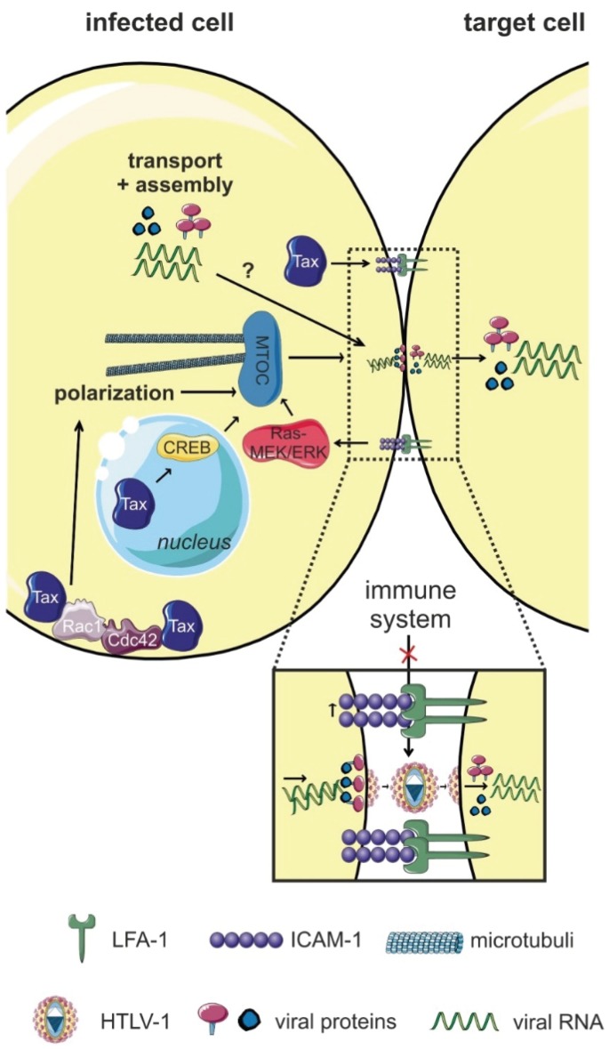
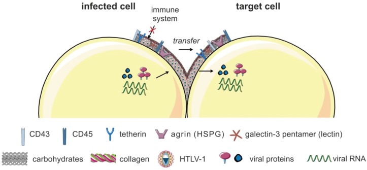
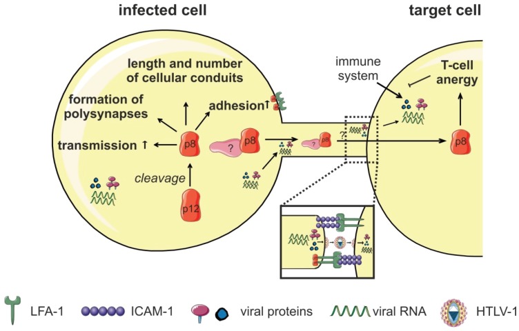
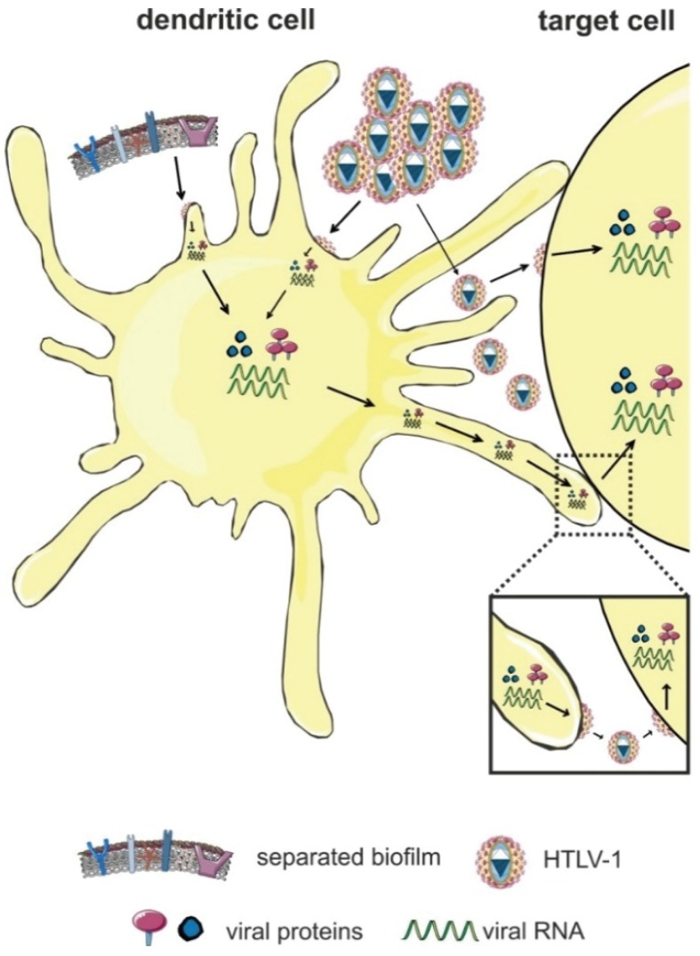
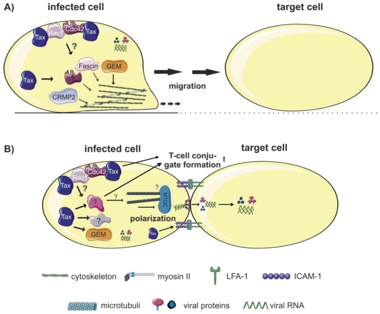
The tumorvirus human T-cell lymphotropic virus type 1 (HTLV-1), a member of the delta-retrovirus family, is transmitted via cell-containing body fluids such as blood products, semen, and breast milk. In vivo, HTLV-1 preferentially infects CD4⁺ T-cells, and to a lesser extent, CD8⁺ T-cells, dendritic cells, and monocytes. Efficient infection of CD4⁺ T-cells requires cell-cell contacts while cell-free virus transmission is inefficient. Two types of cell-cell contacts have been described to be critical for HTLV-1 transmission, tight junctions and cellular conduits. Further, two non-exclusive mechanisms of virus transmission at cell-cell contacts have been proposed: (1) polarized budding of HTLV-1 into synaptic clefts; and (2) cell surface transfer of viral biofilms at virological synapses. In contrast to CD4⁺ T-cells, dendritic cells can be infected cell-free and, to a greater extent, via viral biofilms in vitro. Cell-to-cell transmission of HTLV-1 requires a coordinated action of steps in the virus infectious cycle with events in the cell-cell adhesion process; therefore, virus propagation from cell-to-cell depends on specific interactions between cellular and viral proteins. Here, we review the molecular mechanisms of HTLV-1 transmission with a focus on the HTLV-1-encoded proteins Tax and p8, their impact on host cell factors mediating cell-cell contacts, cytoskeletal remodeling, and thus, virus propagation.

Keywords: HTLV-1; Tax; cell-cell contacts; cell-to-cell transmission; cellular conduit; p8; viral biofilm; virological synapse; virus transmission.


Figures

Figure 1
Figure 1
The virological synapse (VS). Interactions of intercellular adhesion molecule 1 (ICAM-1; on HTLV-1-infected T-cells) with lymphocyte function-associated antigen (LFA-1; on target cells), and signals induced by the viral Tax protein trigger polarization of the microtubule organizing center (MTOC) towards the cell-cell contact and formation of the VS at the cell-cell contact. Tax is not only located in the nucleus, but also at the MTOC and in the cell-cell contact region. Tax-induced CREB signaling (nuclear activity of Tax), the accumulation of Tax at the MTOC, and ICAM-1-induced Ras/MEK/ERK signaling are important for MTOC polarization. It is assumed that the VS allows for efficient polarized budding and virus transmission via a synaptic cleft, thus, avoiding recognition of HTLV-1 by the host immune system. Figure was realized thanks to Servier Medical Art.
Figure 2
Figure 2
The viral biofilm. HTLV-1 virions are accumulated in a specialized extracellular matrix (ECM), the so-called viral biofilm, on the surface of infected cells. The viral biofilm is composed of carbohydrates, components of the ECM (collagen, agrin), linker proteins (galectin-3, tetherin), and O-glycosylated surface receptors (CD43, CD45). HTLV-1 particles are concentrated into large, highly infectious assemblies that cluster towards the cell-cell contact. HTLV-1 is transferred to target cells and guarded by the biofilm from immune recognition. Figure was realized thanks to Servier Medical Art.
Figure 3
Figure 3
Cellular conduits. The viral accessory protein p12 is proteolytically cleaved into the p8 protein, which increases adhesion of T-cells through lymphocyte function-associated antigen-1 (LFA-1) clustering. Further, p8 induces polysynapse formation and enhances the number and length of cellular conduits between T-cells, thereby, enhancing HTLV-1-transmission. p8 is transferred to target cells through these conduits and it is hypothesized to induce T-cell anergy in the target cell. This might be a strategy for HTLV-1 to evade the host’s immune surveillance during infection. Host cell proteins that interact with p8 to enhance conduit formation, p8 transfer, and HTLV-1 transmission are still unknown. Figure was realized thanks to Servier Medical Art.
Figure 4
Figure 4
Transmission of HTLV-1 via dendritic cells (DC). DC either capture the virus and transmit it to target cells in the absence of infection (trans-infection), or they are productively-infected before viral transmission (cis-infection). Productive cell-free infection of DC is achieved in vitro by highly-concentrated preparations of cell-free HTLV-1 or by viral biofilms. Figure was realized thanks to Servier Medical Art.
Figure 5
Figure 5
Host factors regulating cellular migration, invasion and conjugate formation. (A) Proteins enhancing cellular migration and/or invasion of HTLV-1-infected cells could favor dissemination of HTLV-1 to target cells. Expression of the Tax-induced small GTP-binding protein GEM enhances both migration of HTLV-1-infected cells and viral transmission. Activity of CRMP2, a phosphoprotein involved in cytoskeleton rearrangement, is modulated by Tax and correlates with migration of infected cells. The actin-bundling protein Fascin is induced by Tax and important for invasive migration of HTLV-1-infected cells. A role of CRMP2 and Fascin for viral transmission remains to be determined. Both Rac-1 and Cdc42 are interaction partners of Tax that are crucial for migration and for MTOC polarization. (B) T-cell conjugate formation, a prerequisite for cell-to-cell transmission depends on components of the cytoskeleton like the Tax-inducible GEM protein, and on Rac1 and Cdc42. Additionally, Tax regulates expression of surface receptors (see Table 1), which are important for cell-cell contact formation, and, potentially, for formation of the VS and HTLV-1 transmission. The influence of different host factors on polarized budding and formation of the VS remains to be determined. Figure was realized thanks to Servier Medical Art.

Similar articles

Cited by

References

    1. Poiesz B.J., Ruscetti F.W., Gazdar A.F., Bunn P.A., Minna J.D., Gallo R.C. Detection and isolation of type C retrovirus particles from fresh and cultured lymphocytes of a patient with cutaneous T-cell lymphoma. Proc. Natl. Acad. Sci. USA. 1980;77:7415–7419. doi: 10.1073/pnas.77.12.7415. - DOI - PMC - PubMed
    1. Yoshida M., Seiki M., Yamaguchi K., Takatsuki K. Monoclonal integration of human T-cell leukemia provirus in all primary tumors of adult T-cell leukemia suggests causative role of human T-cell leukemia virus in the disease. Proc. Natl. Acad. Sci. USA. 1984;81:2534–2537. doi: 10.1073/pnas.81.8.2534. - DOI - PMC - PubMed
    1. Yoshida M., Miyoshi I., Hinuma Y. Isolation and characterization of retrovirus from cell lines of human adult T-cell leukemia and its implication in the disease. Proc. Natl. Acad. Sci. USA. 1982;79:2031–2035. doi: 10.1073/pnas.79.6.2031. - DOI - PMC - PubMed
    1. Osame M., Usuku K., Izumo S., Ijichi N., Amitani H., Igata A., Matsumoto M., Tara M. HTLV-I associated myelopathy, a new clinical entity. Lancet. 1986;1:1031–1032. doi: 10.1016/S0140-6736(86)91298-5. - DOI - PubMed
    1. Gessain A., Barin F., Vernant J.C., Gout O., Maurs L., Calender A., de The G. Antibodies to human T-lymphotropic virus type-I in patients with tropical spastic paraparesis. Lancet. 1985;2:407–410. doi: 10.1016/S0140-6736(85)92734-5. - DOI - PubMed

Publication types

MeSH terms