. 2016 Mar 23:6:23616.
doi: 10.1038/srep23616.

TEX101, a glycoprotein essential for sperm fertility, is required for stable expression of Ly6k on testicular germ cells

Affiliations

TEX101, a glycoprotein essential for sperm fertility, is required for stable expression of Ly6k on testicular germ cells

Shuichiro Endo et al. Sci Rep. .

Abstract

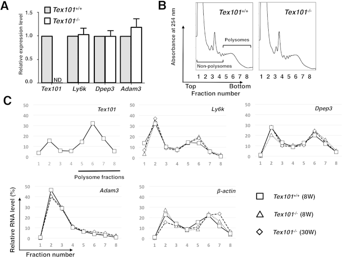
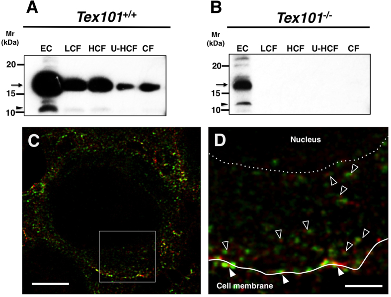
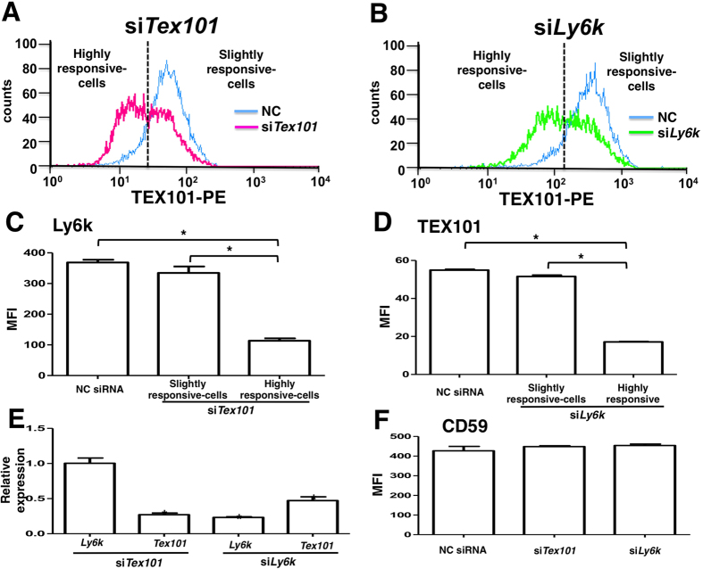
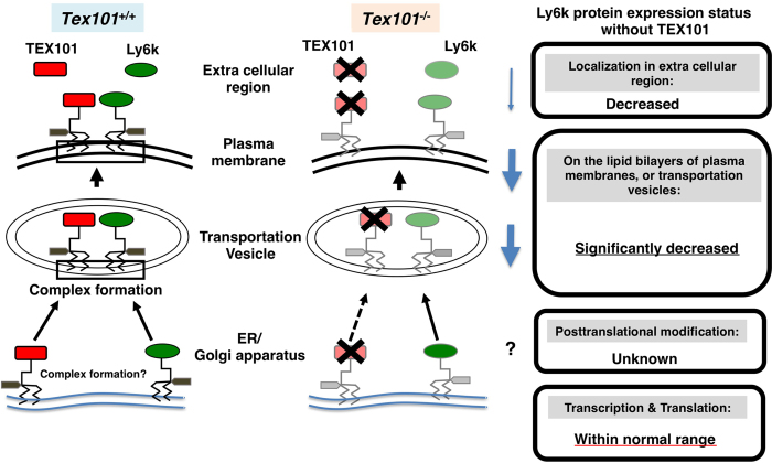
TEX101, a germ cell-specific glycosyl-phosphatidylinositol (GPI)-anchored glycoprotein, is associated with Ly6k during spermatogenesis in testis. Although both Tex101(-/-) and Ly6k(-/-) mice can produce morphologically intact spermatozoa, both knockout mice show an infertile phenotype due to a disorder of spermatozoa to migrate into the oviduct. Since Ly6k specifically interacts with TEX101, complex formation of TEX101/Ly6k appears to be potentially important for functional sperm production. This study evaluated the fate of Ly6k in the presence or absence of TEX101 to explore the molecular interaction of both GPI-anchored proteins in seminiferous tubules. The present study showed that: 1) Although Ly6k mRNA was detected, the protein was present at very low levels in mature testes of Tex101(-/-) mice, 2) Ly6k mRNA level was within the normal range in Tex101(-/-) mice, 3) Ly6k mRNA was translated into a polypeptide in the testes of Tex101(+/+) and Tex101(-/-) mice, and 4) TEX101, as well as Ly6k, are co-factors that affect to molecular expression. These results indicate that both TEX101 and Ly6k contribute to the post-translational counterpart protein expression at the cell membrane. This mechanism may be important in maintaining the production of fertile spermatozoa during spermatogenesis.


Figures

Figure 1
Figure 1. Differences of testicular gene expression between Tex101+/+ and Tex101−/−mice.
Analysis of Tex101, Ly6k, Dpep3, and Adam3 mRNA expression in the Tex101−/− mice (A). Levels of these mRNA expressions in the testes of Tex101+/+ (n = 3) and Tex101−/−(n = 4) mice were measured using qRT-PCR with SYBR Green method. To obtain the ΔΔCt values for the calculation of fold increases, β-actin mRNA was used as a quantitative internal control. Average of each mRNA expression level in Tex101+/+ mouse testis was defined as “relative expression value = 1.0”. The data are indicated with SE. ND: not detectable. Polysome profiles (254 nm absorbance profile) of sucrose gradient sedimentation of cell extracts from the Tex101+/+ and Tex101−/−mice testes (B). Detergent-treated cell extracts were fractionated over a 15–60% sucrose density gradient into eight fractions of equal volume. Expression levels of testicular Tex101, Ly6k, Dpep3, Adam3, and β-actin mRNAs from the 8-week Tex101+/+ and 8- or 30-week-old Tex101−/−mice in each fraction of the sucrose gradients (C). Each fraction number corresponds to that in the absorbance profile shown in (B). Data are graphically represented as ratio of transcripts from a typical experiment, present in each fraction to the sum of all fractions. As an external standard, r-luc mRNA was used.
Figure 2
Figure 2. Effects of Tex101 gene disruption on the expression of testicular proteins.
Western blot analysis (A). The EC, WS, and TS fractions of testicular proteins from Tex101+/+ or Tex101−/− mice were applied to each lane of a 15% SDS-PAGE gel, separated under non-reducing (TEX101) or reducing (Ly6k and DPEP3) conditions. After electroblotting, the immunoreactivity of the Abs against TEX101, DPEP3, or Ly6k was monitored with an ECL detection system. Mr: molecular mass. Arrows indicate the main immunoreactive bands detected with each specific Ab. A non-membrane-binding form of Ly6k at the apparent molecular mass of 12-kDa is indicated by an arrow head. Immunofluorescent analysis (B). PFA-fixed frozen sections of the Tex101+/+ or Tex101−/− mouse testis were incubated with the anti-TEX101, DPEP3, or Ly6k Abs, or isotype-matched NC Abs corresponding to each primary Ab (green), and following nuclear staining with DAPI (blue). All seminiferous tubules shown are stage VIII based on nuclear staining. Bars: 50 μm.
Figure 3
Figure 3. Subcellular distributions of Ly6k in the testes derived from TEX101+/+ and Tex101−/−mice.
Western blot analyses of extracellular and testicular-cell fractions (A,B). The testicular-cell fractions prepared with the differential centrifugation method as described “Materials and Methods” section. The samples were adjusted to same cell numbers in equal volume (10 μl), applied to each lane of 15% SDS-PAGE gels, electroblotted onto a PVDF membranes, and then probed with the anti-Ly6k pAb. EC: extracellular fraction; LCF: fraction by low-speed centrifugation; HCF: fraction by high-speed centrifugation; U-HCF: fraction by ultrahigh-speed centrifugation; and CF: cytosol fraction. Apparent molecular mass (Mr) of Ly6k is indicated by an arrowhead at the left margin of each panel. Immunofluorescent analyses of TEX101 and Ly6k within the seminiferous tubules of Tex101+/+ mouse (C,D). Testicular cryosections (3 μm in thickness) were incubated with the primary antibodies at 4 °C overnight and subsequently with Alexa Fluor 488 or Alexa Fluor 555-conjugated secondary Abs. The specimens were observed by a TCS SP8 STED 3X confocal laser microscopy. The localizations of TEX101 and Ly6k are indicated as red and green, respectively. An overlay image is showed in C. Panel (D) shows a higher magnified image of the boxed region in panel (C). Co-localization of TEX101 and Ly6k on the plasma membranes and in cytoplasmic regions is indicated with closed and open arrowheads, respectively. A solid line in D indicates cell surface of a TGC, and a dotted line represent a border between cytoplasm and nucleus of the cell. Bars: 5 μm (C); 1 μm (D).
Figure 4
Figure 4. Effects of siRNAs against Tex101 and Ly6k genes on the expression of those molecules in 293mTx/ly6kκB cell line.
After transfected with each siRNA and sequentially incubated for 48 h in a 5% CO2 humidified atmosphere at 37 °C, the 293mTx/ly6kκB cells were harvested. Expression levels of TEX101 and Ly6k on the transfected cell surfaces were measured by flow cytometry. Histograms of the cells transfected either Tex101 (A), or Ly6k (B) siRNA. Blue lines represent the histogram of NC siRNA transfected cells. The left sides peak of dotted lines contain cells that are highly responsive for siTex101 (A) or siLy6k (B), respectively. The other side peaks contain cells that are slightly response for each siRNA. Expression of Ly6k (C) and TEX101 (D) in the 293mTx/ly6kκB cells transfected with NC, Tex101, or Ly6k siRNA. The data are expressed as the mean fluorescence intensity (MFI) with SE measured by flow cytometry (n = 3). *p < 0.05. Changes in mRNA expression levels of Ly6k and Tex101 in the TEX101- or Ly6k siRNA-transfected 293mTx/ly6kκB cells (E). The mRNA expression levels were measured by the qRT-PCR analysis with SYBR Green method. Relative expression values were normalized with the Rps18 expression and the normalized values of each mRNA expression in the transfected cells with NC siRNA are defined as “1.0”. Values are shown as means with SE (n = 3). *p < 0.05 compared to the control (NC siRNA transfection). Effects of Tex101 and Ly6k siRNAs on CD59 expression (F). After 293mTx/ly6kκB cells were transfected with NC siRNA, siTex101, or siLy6k and then incubated, CD59 expression levels were analyzed by flow cytometry. The data shown are the MFI with SE (n = 3).
Figure 5
Figure 5. Molecular status of Ly6k with/without TEX101 in the TGCs.
The left diagram indicates TEX101/Ly6k complex formation of wild-type (Tex101+/+) mouse. After translation, GPI remodeling of these molecules is completed from ER to Golgi apparatus, then these molecules are expressed as TEX101/Ly6k complex (represented by black square) on lipid bilayers including transportation vesicle and plasma membrane. In addition, (a part of) both TEX101 and Ly6k are released into extracellular space. In TEX101-null TGCs (the right diagram), Ly6k expression is drastically decreased. Black cross marks indicate the disruption of the molecules. The potential status of Ly6k protein expression without TEX101 is boxed.

References

    1. Russell L. D. et al. Mammalian spermatogenesis in Histological and histopathological evaluation of the testis (ed. Russell L. D. et al.) 1–40 (Cache River Press, 1990).
    1. Bowles J. & Koopman P. Sex determination in mammalian germ cells: extrinsic versus intrinsic factors. Reproduction 139, 943–958 (2010). - PubMed
    1. Kurita A. et al. Identification, cloning, and initial characterization of a novel mouse testicular germ cell-specific antigen. Biol Reprod 64, 935–945 (2001). - PubMed
    1. Takayama T. et al. Sexually dimorphic expression of the novel germ cell antigen TEX101 during mouse gonad development. Biol Reprod 72, 1315–1323 (2005). - PubMed
    1. Jin H. et al. Molecular characterization of a germ-cell-specific antigen, TEX101, from mouse testis. Zygote 14, 201–208 (2006). - PubMed

Publication types