. 2016 May 6;291(19):9872-81.
doi: 10.1074/jbc.M116.720029. Epub 2016 Mar 22.

Brain-derived Neurotrophic Factor in Megakaryocytes

Affiliations

Brain-derived Neurotrophic Factor in Megakaryocytes

Pedro Chacón-Fernández et al. J Biol Chem. .

Abstract

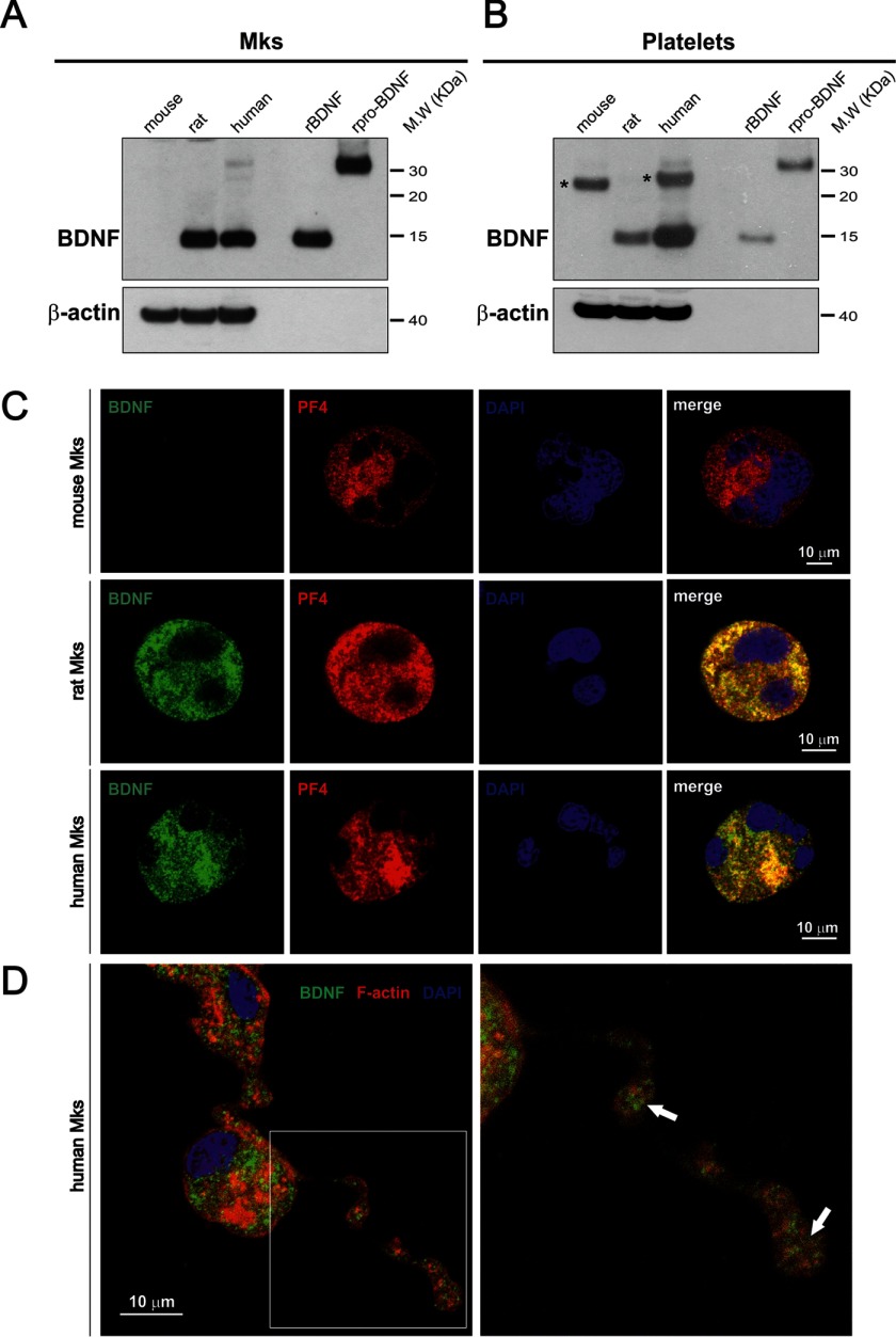
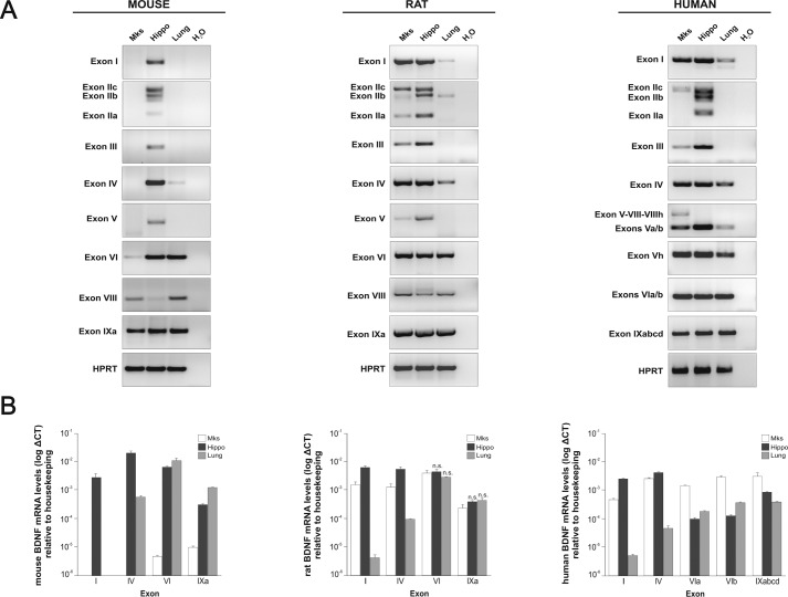
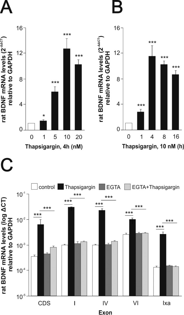
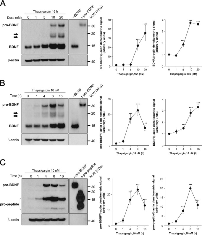
The biosynthesis of endogenous brain-derived neurotrophic factor (BDNF) has thus far been examined in neurons where it is expressed at very low levels, in an activity-dependent fashion. In humans, BDNF has long been known to accumulate in circulating platelets, at levels far higher than in the brain. During the process of blood coagulation, BDNF is released from platelets, which has led to its extensive use as a readily accessible biomarker, under the assumption that serum levels may somehow reflect brain levels. To identify the cellular origin of BDNF in platelets, we established primary cultures of megakaryocytes, the progenitors of platelets, and we found that human and rat megakaryocytes express the BDNF gene. Surprisingly, the pattern of mRNA transcripts is similar to neurons. In the presence of thapsigargin and external calcium, the levels of the mRNA species leading to efficient BDNF translation rapidly increase. Under these conditions, pro-BDNF, the obligatory precursor of biologically active BDNF, becomes readily detectable. Megakaryocytes store BDNF in α-granules, with more than 80% of them also containing platelet factor 4. By contrast, BDNF is undetectable in mouse megakaryocytes, in line with the absence of BDNF in mouse serum. These findings suggest that alterations of BDNF levels in human serum as reported in studies dealing with depression or physical exercise may primarily reflect changes occurring in megakaryocytes and platelets, including the ability of the latter to retain and release BDNF.

Keywords: blood; brain-derived neurotrophic factor (BDNF); neurobiology; neurochemistry; neurotrophin.


Figures

FIGURE 1.
FIGURE 1.
Differential BDNF protein levels in mouse, rat, and human megakaryocytes and platelets. Western blot lysates of cultured Mks (A) and blood platelets (B) are shown. Eighty micrograms of protein per lane were loaded, and the blotting membrane was incubated with the mouse monoclonal antibody 3C11 developed by Icosagen (Tartu, Estonia). Recombinant BDNF and pro-BDNF were used as molecular mass markers and antibodies to β-actin as loading controls. Asterisks (top right panels) point to a band unrelated to BDNF likely corresponding to immunoglobulin light chains in the mouse sample. Note the absence of BDNF in mouse Mks and platelets. C, antibodies to BDNF 9 (green) (15) and PF4 (red) reveal expression of both antigens in mature rat and human Mks. Note that unlike PF4, BDNF is not detectable in mouse Mks. The co-localization of BDNF with PF4 in rat and human Mks was quantified using the pixel intensity specifically generated by each channel. In humans, 83% and in rats 86% BDNF-positive granules were also PF4-positive. Blue, DAPI staining. D, immunofluorescence staining of F-actin (red) and BDNF (green) in proplatelet-forming cultured human Mks. Arrows indicate BDNF accumulation in proplatelet buds.
FIGURE 2.
FIGURE 2.
Transcriptional analysis of BDNF in mouse, rat, and human megakaryocytes. Conventional (A) and real time quantitative (B) PCR using exon-specific primers with RNA extracted from mature cultured Mks, adult hippocampus (Hippo), and lung are shown. Note that in the mouse, the neuron-specific transcripts, including exon I and IV, are not detected and that by contrast the transcript pattern resembles the non-neuronal pattern observed in lung tissue. The converse is the case with RNA extracted from rat and human Mks with transcript patterns, including exon I and IV, that are characteristic of a neuronal pattern as illustrated with the hippocampus. Unless indicated as non-significant (n.s.), all values are mean values ± S.E. in triplicates and based on three independent experiments, at p < 0.001 (paired t test).
FIGURE 3.
FIGURE 3.
Up-regulation of Bdnf mRNA by thapsigargin. Effect of extracellular calcium. Dose response (A) and time course (B) of Bdnf mRNA expression by rat Mks after thapsigargin treatment. Purified mature rat Mks were cultured in the presence or absence of thapsigargin or vehicle (DMSO) used at the indicated concentrations (A) and for different lengths of times (B). Total mRNA was extracted and reverse-transcribed, and the resulting cDNA was amplified by real time quantitative PCR using specific primers for the coding sequence of Bdnf. C, extracellular calcium dependence of thapsigargin-induced Bdnf mRNA increase. Rat Mks were preincubated with 2.5 mm EGTA for 1.5 h at 37 °C followed by 10 nm thapsigargin for 4 h. mRNA expression was analyzed by real time quantitative PCR using specific primers for the coding sequence of Bdnf (CDS) or exon-specific primers. All values are mean values ± S.E. in triplicates and based on three independent experiments. Unless indicated, all the statistical values are compared with the control. *, p < 0.05; ***, p < 0.001 (paired t test).
FIGURE 4.
FIGURE 4.
Effect of thapsigargin on pro-BDNF, mature BDNF, and pro-peptide in rat Mks. Dose response (A) and time course (B) of pro-BDNF and mature BDNF proteins by rat Mks after thapsigargin treatment are shown. Mature Mks were cultured for 16 h at the indicated doses of thapsigargin (A) or 10 nm thapsigargin for the indicated times (B). Forty micrograms of protein per lane were loaded, and the blotting membrane was incubated with the mouse monoclonal antibody 3C11 developed by Icosagen. Arrows indicate intermediate proteolytic products of pro-BDNF (C). Time course of pro-BDNF and pro-peptide proteins generated by rat Mks incubated with 10 nm thapsigargin for the indicated time periods. Eighty micrograms protein per lane were loaded, and the blotting membrane was incubated with the mouse monoclonal antibody H1001G developed by GeneCopeia, Inc. The blots shown are representative of three independent experiments with similar results. Graphs show mean ± S.E. of the densitometric values quantified from the blots of the three separate experiments. ***, p < 0.001 (paired t test compared the corresponding controls). Recombinant BDNF (150–300 pg), cleavage-resistant recombinant pro-BDNF (0.5–1 ng), and recombinant pro-peptide (1–10 ng) were used as molecular mass markers and antibodies to β-actin as loading controls.
FIGURE 5.
FIGURE 5.
Differential expression of proprotein convertases in primary MKs. Conventional (A) and real time quantitative (B) PCRs using specific primers and RNA extracted from mature cultured rat Mks and adult hippocampus are shown. Note that although transcripts, including Pcsk1, PcsK2, Pcsk4, and Pcsk5, are expressed in hippocampal tissue, they are not detected in Mks (A). Comparative expression levels between the two tissues for the expressed proprotein convertases are shown in B. All values are mean values ± S.E. in triplicates and are based on three independent experiments.

References

    1. Park H., and Poo M. M. (2013) Neurotrophin regulation of neural circuit development and function. Nat. Rev. Neurosci. 14, 7–23 - PubMed
    1. Zagrebelsky M., and Korte M. (2014) Form follows function: BDNF and its involvement in sculpting the function and structure of synapses. Neuropharmacology 76, 628–638 - PubMed
    1. Egan M. F., Kojima M., Callicott J. H., Goldberg T. E., Kolachana B. S., Bertolino A., Zaitsev E., Gold B., Goldman D., Dean M., Lu B., and Weinberger D. R. (2003) The BDNF val66met polymorphism affects activity-dependent secretion of BDNF and human memory and hippocampal function. Cell 112, 257–269 - PubMed
    1. Gray J., Yeo G. S., Cox J. J., Morton J., Adlam A. L., Keogh J. M., Yanovski J. A., El Gharbawy A., Han J. C., Tung Y. C., Hodges J. R., Raymond F. L., O'rahilly S., and Farooqi I. S. (2006) Hyperphagia, severe obesity, impaired cognitive function, and hyperactivity associated with functional loss of one copy of the brain-derived neurotrophic factor (BDNF) gene. Diabetes 55, 3366–3371 - PMC - PubMed
    1. Leibrock J., Lottspeich F., Hohn A., Hofer M., Hengerer B., Masiakowski P., Thoenen H., and Barde Y. A. (1989) Molecular cloning and expression of brain-derived neurotrophic factor. Nature 341, 149–152 - PubMed

Publication types

LinkOut - more resources