. 2016 Apr 5;113(14):3785-90.
doi: 10.1073/pnas.1600211113. Epub 2016 Mar 22.

A heme-binding domain controls regulation of ATP-dependent potassium channels

Affiliations

A heme-binding domain controls regulation of ATP-dependent potassium channels

Mark J Burton et al. Proc Natl Acad Sci U S A. .

Abstract

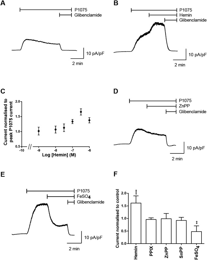
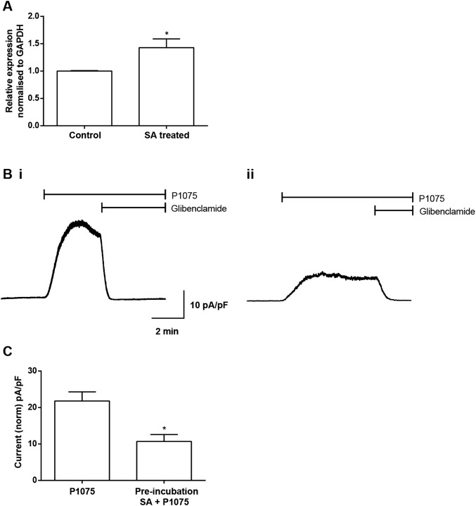
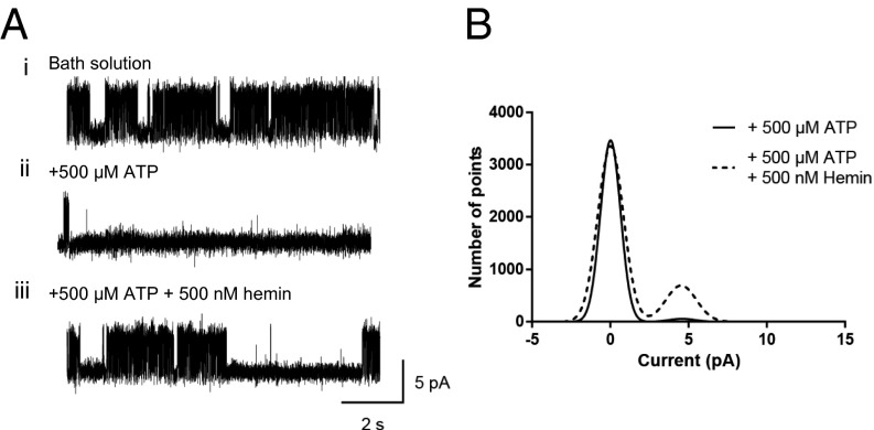
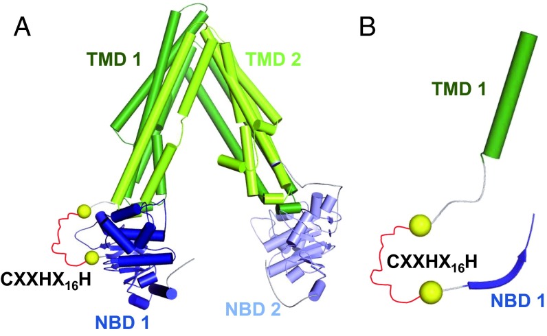
Heme iron has many and varied roles in biology. Most commonly it binds as a prosthetic group to proteins, and it has been widely supposed and amply demonstrated that subtle variations in the protein structure around the heme, including the heme ligands, are used to control the reactivity of the metal ion. However, the role of heme in biology now appears to also include a regulatory responsibility in the cell; this includes regulation of ion channel function. In this work, we show that cardiac KATP channels are regulated by heme. We identify a cytoplasmic heme-binding CXXHX16H motif on the sulphonylurea receptor subunit of the channel, and mutagenesis together with quantitative and spectroscopic analyses of heme-binding and single channel experiments identified Cys628 and His648 as important for heme binding. We discuss the wider implications of these findings and we use the information to present hypotheses for mechanisms of heme-dependent regulation across other ion channels.

Keywords: KATP channel; SUR2A; heme; heme regulation; potassium channel.


Conflict of interest statement

The authors declare no conflict of interest.

Figures

Fig. S1.
Fig. S1.
(A) Schematic representation of cardiac sarcolemmal KATP channel structure; four SUR2A subunits assemble with four pore-lining Kir6.2 subunits to form an octameric channel structure. Highlighted in red is the disordered protein region which contains the motif CXXHX16H leading into nucleotide binding domain 1 (NBD1). (B) Multiple sequence alignment of the human ATP-binding cassette subfamily C (CFTR/MRP) proteins using the CLUSTAL color scheme. Shown is the section of the alignment covering the CXXHX16H motif and parts of the adjacent transmembrane domain 1 (TMD1) and nucleotide binding domain 1 (NBD1). The SUR2A rat sequence was added for comparison.
Fig. S2.
Fig. S2.
Heme increases cardiac KATP currents. (A) Whole-cell recording of KATP currents (current amplitudes are normalized to cell capacitance and expressed as pA/pF) from an isolated ventricular myocyte. Currents were recorded at 0 mV, and P1075 (10 µM) and glibenclamide (10 µM) were bath applied as indicated by the bar. (B) Whole-cell recording of KATP currents, as above, with addition of hemin (500 nM) as indicated by the bar. (C) Concentration response curve for hemin-induced increase in the KATP current in isolated ventricular myocytes. Experiments were repeated, for the dataset for log [hemin] −9, −8, and −7.5, n = 6; −7 and −6.5, n = 7; and −6, n = 8. Whole-cell recordings of KATP currents from isolated ventricular myocytes from a holding potential of 0 mV, and P1075 (10 µM), with (D) zinc protoprorphyrin IX (ZnPP) (500 nM) or (E) FeSO4 (500 nM) and glibenclamide (10 µM), were bath applied as indicated by the bar. (F) Summary graph showing the change in KATP current normalized to P1075 current of ventricular myocytes either untreated (control = 1) (n = 6) or treated with hemin (n = 7), protoporphyrin IX (PP) (n = 3) zinc protoprorphyrin IX (ZnPP) (n = 3), tin protophorphyrin IX (SnPP) (n = 4), and FeSO4 (n = 5). ***P ≤ 0.001, **P ≤ 0.01 vs. control. ANOVA, Dunnett’s post hoc test.
Fig. S3.
Fig. S3.
Inhibition of heme synthesis causes a reduction in KATP currents recorded from adult rat ventricular myocytes. (A) qPCR, normalized to the housekeeping gene GAPDH, showed an up-regulation of ALAS-1 in response to inhibition of heme synthesis with SA (1 mM for 4 h). *P ≤ 0.05, n = 4, significantly different from control with Student t test. (B, i) Representative whole-cell trace of normalized KATP current (pA/pF) in response to P1075 (10 μM) compared with (B, ii) a current recording from myocytes pretreated with SA (1 mM) for 4 h. (C) Mean peak current elicited by P1075 for control myocytes and those incubated in SA for 4 h illustrated a significant decrease from 21.8 ± 2.5, n = 28, to 10.7 ± 1.8, n = 8 (unpaired t test, *P < 0.05).
Fig. 1.
Fig. 1.
Heme increases KATP single channel open probabilities. (A, i) KATP channel currents recorded from an inside-out patch of a ventricular myocyte at +70 mV exposed to normal bath solution containing 0 μM ATP, illustrating high channel activity. (A, ii) Local perfusion of the same patch with 500 µM ATP reduced channel activity. (A, iii) Local perfusion of the same patch with 500 µM ATP and 500 nM hemin increased channel activity. (B) Overlaid amplitude histograms from the above patch, (fitted with Gaussian distributions) illustrating an increase in single channel activity with application of 500 nM hemin (dashed line).
Fig. 2.
Fig. 2.
(A) SUR2A homology model based on C. elegans MRP PGP-1. α-Helices are displayed as barrels, and β-strands as arrows using the color scheme introduced in Fig. S1A. The CXXHX16H motif (red), which could not be modeled, is schematically indicated as a stretch of residues between the two yellow spheres. (B) Zoom into the SUR2A homology model. Visualization as in A, but only residues that are also present in the multiple sequence alignment in Fig. S1B are shown.
Fig. 3.
Fig. 3.
Effect of mutagenesis of the CXXHX16H region of SUR2A on KATP channel activity. (A) Inside-out patch of WT perfused with 500 µM ATP, and 500 µM ATP with 500 nM hemin, and (B) plot mean Popen with empty bars indicating 500 µM ATP and gray bars indicating 500 µM ATP with 500 nM hemin, n = 9 (***P ≤ 0.001). (C) Representative inside-out patch of C628S with 500 µM ATP, and 500 µM ATP with 500 nM hemin, and (D) mean Popen, n = 7. (E) Representative inside-out patch of H648A with 500 µM ATP, and 500 µM ATP with 500 nM hemin, and (F) mean Popen for 500 µM ATP and 500 µM ATP with 500 nM hemin, n = 5. (G) Mean Popen for all mutants: empty bars for 500 µM ATP and gray bars indicating 500 µM ATP with 500 nM hemin.
Fig. S4.
Fig. S4.
Heterologously expressed KATP channels are activated by heme. (A) Patches were exposed to 0 µM ATP solution (i), locally perfused with 500 µM ATP (ii), and 500 µM ATP plus 500 nM hemin (iii). Exposure to the KATP blocker glibenclamide (50 µM) inhibited channel activity (iv). (B) Popen values are plotted and (C) mean increases in Popen are observed from 0.023 ± 0.009, n = 9, for +500 µM ATP alone to 0.109 ± 0.020, n = 9 (***P ≤ 0.001), for +500 ATP + 500 nM hemin. (D) Macropatch recordings from KATP channels illustrate increase in channel current at all potentials with the presence of 500 nM hemin, n = 3. (E) Plots of Popen from inside-out patches of C628S SUR2A mutation perfused with 500 µM ATP and 500 µM ATP with 500 nM hemin with no significant change in activity between 500 µM ATP = 0.053 ± 0.019, n = 7 and 500 µM ATP with 500 nM hemin = 0.043 ± 0.014, n = 7. (F) All Popen values recorded from H648A SUR2A mutation with 500 µM ATP, 500 µM ATP with 500 nM hemin with no significant difference between 500 µM ATP = 0.009 ± 0.005, n = 5, and 500 µM ATP with 500 nM hemin = 0.011 ± 0.008, n = 5.
Fig. 4.
Fig. 4.
Spectroscopic analysis of heme bound to SUR2A and a synthetic peptide. (A) Spectrophotometric titrations of the synthetic peptide LPFESCKKHTGVQSKPINRKQPGRYHLDNYE with heme. (B) Corresponding heme titration of the SUR2A truncated protein (residues S615-L933) containing the heme-binding CXXHX16H region; the arrows represent the directions of the absorbance changes with increasing heme concentration. (C) Room temperature high-frequency resonance Raman spectra of (i) hemin, (ii) ferric SUR2A(S615-L933)-hemin at substoichiometric hemin concentrations, (iii) ferric SUR2A(S615-L933)-hemin with the protein in four- to fivefold excess, and (iv) H648A-hemin. All spectra collected with 413.1-nm laser excitation.
Fig. S5.
Fig. S5.
High-frequency resonance Raman spectra of (i) hemin, (ii) the SUR2A(S615-L933)-hemin immediately after hemin addition, and (iii) the SUR2A(S615-L933)-hemin complex 4 h after hemin addition.
Fig. S6.
Fig. S6.
X-band EPR spectra of (i) hemin, (ii) SUR2A(S615-L933)-hemin complex, (iii) SUR2A(S615-L933)-hemin complex (four- to fivefold excess of SUR2A), (iv) H648A-hemin complex, (v) C628S-hemin complex, and (vi) H631A-hemin complex. The signals indicated by * and ** indicate an artifact signal and minor Cu(II) contamination, respectively. Experimental conditions: microwave frequency, 9.39 GHz; microwave power, 1 (Left) and 0.25 mW (Right); one (Left) and four scans (Right) per spectrum; field modulation amplitude, 2 mT; field modulation frequency, 100 kHz; temperature, 20 K; [heme] = 100 µM.
Fig. S7.
Fig. S7.
Blumberg-Peisach correlation diagram including various heme proteins. The figure shows a correlation between the strength of the axial field in units of the spin−orbit coupling constant λ (x axis) and the ratio of the rhombic and axial crystal field parameters (y axis). SUR2A lies in the region designated for Cys(S)/X heme coordination environment (15).
Fig. 5.
Fig. 5.
Cartoon representation of heme interaction in various ion channels. (A) In the KATP channel (this work), heme binds to the CXXHX16H motif in the unique insertion (G622-P665 in the human protein) on the cytoplasmic domain of SUR2A and increases the open channel probability. (B) In Slo1 (BK) channel (5), heme binds to the cytochrome c-like motif CXXCH in the disordered region between the RCK1 and RCK2 domains, and increased concentrations of heme inhibit K+ currents by decreasing the frequency of channel opening. Inhibition of channel activity as heme concentration increases is consistent with other observations (41) in which knockdown of heme oxygenase (which would increase heme concentration) also reduces channel activity. (C) In the Kv1.4 channel (23), heme binds to the “ball-and-chain” N terminus of the A-type potassium channel and impairs the inactivation process; a heme-responsive CXXHX18H motif is suggested as being responsible for heme binding, which introduces a stable configuration in the otherwise disordered region. The membrane is depicted in pale blue and the intracellular side is on the bottom. The light purple rectangles depict the conduction pore of the inward rectifier K+ channel Kir6.2 subunit in the KATP channel and the Slo1 and Kv1.4 channels. The gray (TMD0), dark green (TMD1), and light green (TMD2) rectangles represent the transmembrane domains of the sulphonylurea receptor SUR2A in the KATP channel (color scheme as in (Fig. S1A), and the dark purple rectangles are the voltage-sensor domains in the Slo1 and Kv1.4 channels. Other transmembrane domains have been omitted for simplicity. NBD, nucleotide binding domains 1 and 2; RCK1/RCK2, regulator of conductance K domains 1 and 2. Heme is depicted as a red diamond.

Similar articles

Cited by

References

    1. Hou S, Reynolds MF, Horrigan FT, Heinemann SH, Hoshi T. Reversible binding of heme to proteins in cellular signal transduction. Acc Chem Res. 2006;39(12):918–924. - PubMed
    1. Rodgers KR. Heme-based sensors in biological systems. Curr Opin Chem Biol. 1999;3(2):158–167. - PubMed
    1. Shimizu T. Binding of cysteine thiolate to the Fe(III) heme complex is critical for the function of heme sensor proteins. J Inorg Biochem. 2012;108:171–177. - PubMed
    1. Smith AG, Raven EL, Chernova T. The regulatory role of heme in neurons. Metallomics. 2011;3(10):955–962. - PubMed
    1. Shimizu T, et al. Gaseous O2, NO, and CO in signal transduction: Structure and function relationships of heme-based gas sensors and heme-redox sensors. Chem Rev. 2015;115(13):6491–6533. - PubMed

Publication types

LinkOut - more resources