

Hybrid Discrete Wavelet Transform and Gabor Filter Banks Processing for Features Extraction from Biomedical Images
- PMID: 27006906
- PMCID: PMC4782617
- DOI: 10.1155/2013/104684
Hybrid Discrete Wavelet Transform and Gabor Filter Banks Processing for Features Extraction from Biomedical Images
Abstract
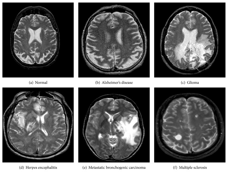
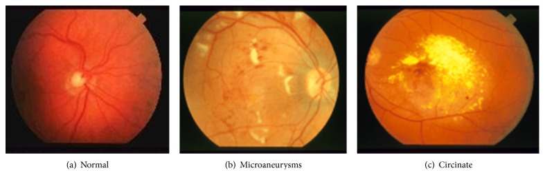
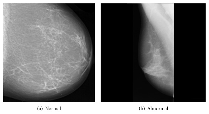
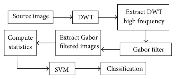
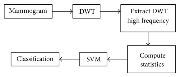
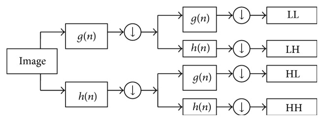
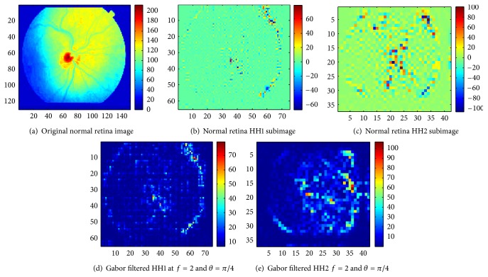
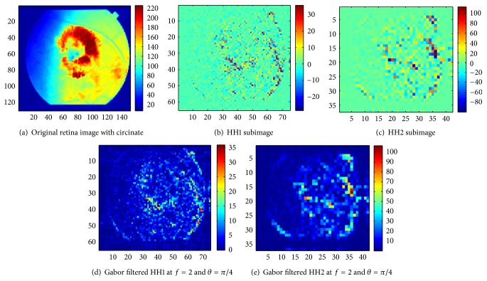
A new methodology for automatic feature extraction from biomedical images and subsequent classification is presented. The approach exploits the spatial orientation of high-frequency textural features of the processed image as determined by a two-step process. First, the two-dimensional discrete wavelet transform (DWT) is applied to obtain the HH high-frequency subband image. Then, a Gabor filter bank is applied to the latter at different frequencies and spatial orientations to obtain new Gabor-filtered image whose entropy and uniformity are computed. Finally, the obtained statistics are fed to a support vector machine (SVM) binary classifier. The approach was validated on mammograms, retina, and brain magnetic resonance (MR) images. The obtained classification accuracies show better performance in comparison to common approaches that use only the DWT or Gabor filter banks for feature extraction.
Figures
Similar articles
-
A Novel Method for Classifying Liver and Brain Tumors Using Convolutional Neural Networks, Discrete Wavelet Transform and Long Short-Term Memory Networks.Sensors (Basel). 2019 Apr 28;19(9):1992. doi: 10.3390/s19091992. Sensors (Basel). 2019. PMID: 31035406 Free PMC article.
-
[Hyperspectral image classification based on 3-D gabor filter and support vector machines].Guang Pu Xue Yu Guang Pu Fen Xi. 2014 Aug;34(8):2218-24. Guang Pu Xue Yu Guang Pu Fen Xi. 2014. PMID: 25474965 Chinese.
-
Coronavirus disease identification using Multi-subband feature analysis in DWT domain.Procedia Comput Sci. 2023;218:574-584. doi: 10.1016/j.procs.2023.01.039. Epub 2023 Jan 31. Procedia Comput Sci. 2023. PMID: 36743796 Free PMC article.
-
Automated diagnosis of mammogram images of breast cancer using discrete wavelet transform and spherical wavelet transform features: a comparative study.Technol Cancer Res Treat. 2014 Dec;13(6):605-15. doi: 10.7785/tcrtexpress.2013.600262. Epub 2013 Aug 31. Technol Cancer Res Treat. 2014. PMID: 24000991 Free PMC article.
-
Fast 2D Complex Gabor Filter With Kernel Decomposition.IEEE Trans Image Process. 2018 Apr;27(4):1713-1722. doi: 10.1109/TIP.2017.2783621. IEEE Trans Image Process. 2018. PMID: 29346090
Cited by
-
Heuristic neural network approach in histological sections detection of hydatidiform mole.J Med Imaging (Bellingham). 2019 Oct;6(4):044501. doi: 10.1117/1.JMI.6.4.044501. Epub 2019 Nov 5. J Med Imaging (Bellingham). 2019. PMID: 31720313 Free PMC article.
-
DIAROP: Automated Deep Learning-Based Diagnostic Tool for Retinopathy of Prematurity.Diagnostics (Basel). 2021 Nov 3;11(11):2034. doi: 10.3390/diagnostics11112034. Diagnostics (Basel). 2021. PMID: 34829380 Free PMC article.
-
Histo-CADx: duo cascaded fusion stages for breast cancer diagnosis from histopathological images.PeerJ Comput Sci. 2021 Apr 27;7:e493. doi: 10.7717/peerj-cs.493. eCollection 2021. PeerJ Comput Sci. 2021. PMID: 33987459 Free PMC article.
-
Fetal Brain Abnormality Classification from MRI Images of Different Gestational Age.Brain Sci. 2019 Sep 12;9(9):231. doi: 10.3390/brainsci9090231. Brain Sci. 2019. PMID: 31547368 Free PMC article.
-
MB-AI-His: Histopathological Diagnosis of Pediatric Medulloblastoma and its Subtypes via AI.Diagnostics (Basel). 2021 Feb 20;11(2):359. doi: 10.3390/diagnostics11020359. Diagnostics (Basel). 2021. PMID: 33672752 Free PMC article.
References
-
- Lee N., Laine A. F., Smith T. R. Learning non-homogenous textures and the unlearning problem with application to drusen detection in retinal images. Proceedings of the 5th IEEE International Symposium on Biomedical Imaging: From Nano to Macro (ISBI '08); May 2008; Paris, France. pp. 1215–1218. - DOI
-
- Dong A., Wang B. Feature selection and analysis on mammogram classification. Proceedings of the IEEE Pacific Rim Conference on Communications, Computers and Signal Processing (PACRIM '09); August 2009; Victoria, BC, Canada. pp. 731–735. - DOI
-
- Tirtajaya A., Santika D. D. Classification of microcalcification using dual-tree complex wavelet transform and support vector machine. Proceedings of the 2nd International Conference on Advances in Computing, Control and Telecommunication Technologies (ACT '10); December 2010; Jakarta, Indonesia. pp. 164–166. - DOI
LinkOut - more resources
Full Text Sources
Other Literature Sources