

Image Segmentation and Analysis of Flexion-Extension Radiographs of Cervical Spines
- PMID: 27006937
- PMCID: PMC4782582
- DOI: 10.1155/2014/976323
Image Segmentation and Analysis of Flexion-Extension Radiographs of Cervical Spines
Abstract
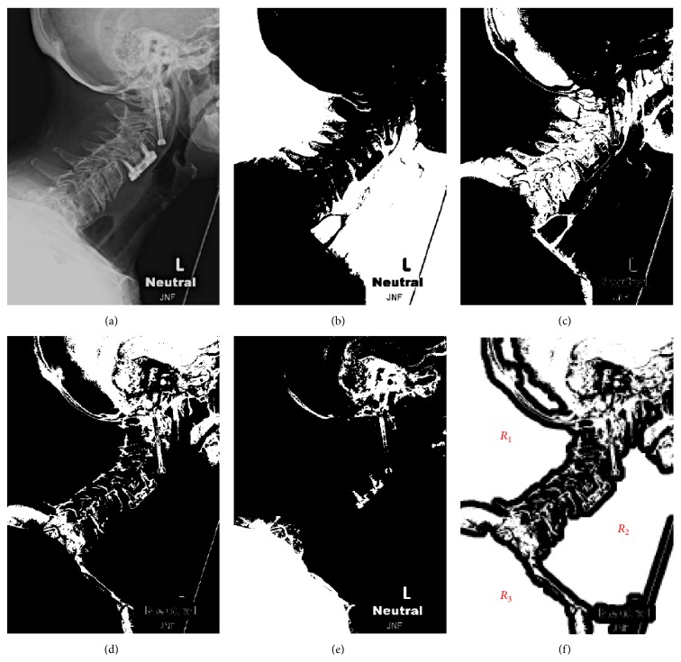
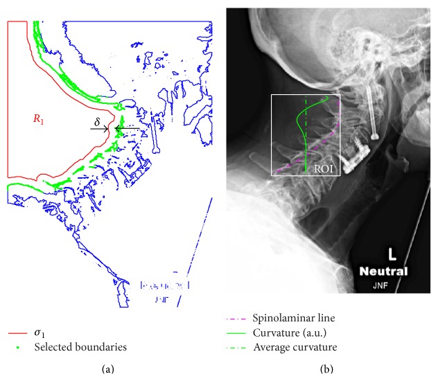
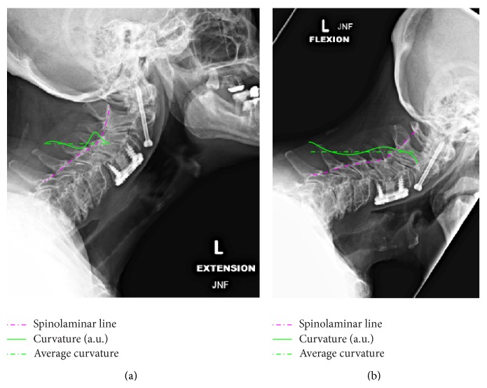
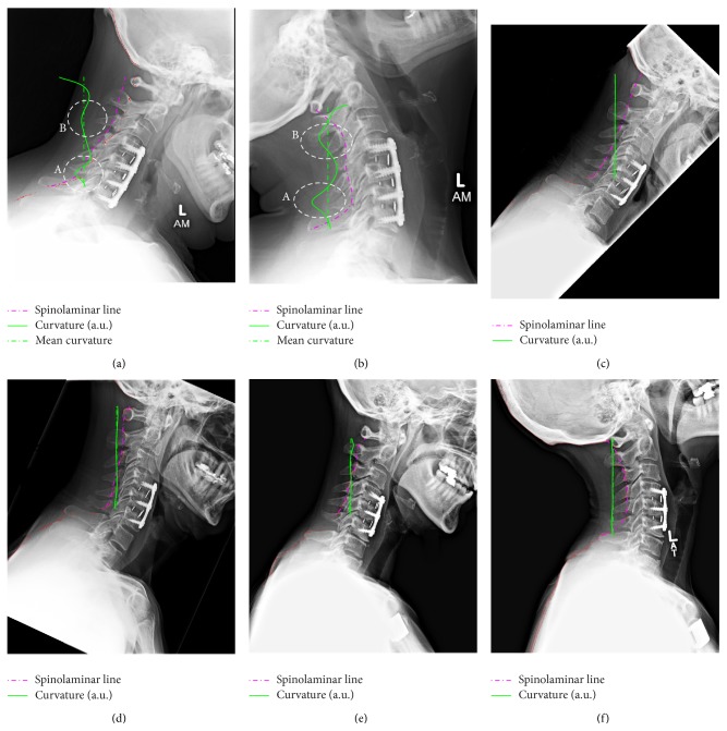
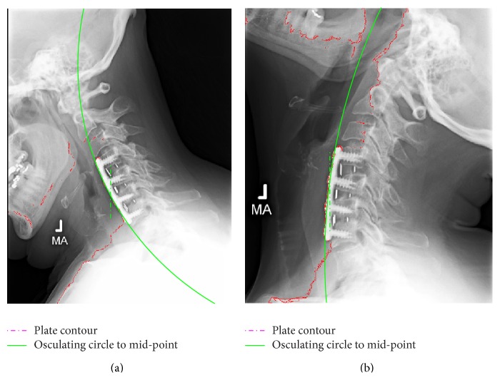
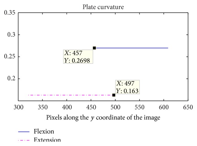
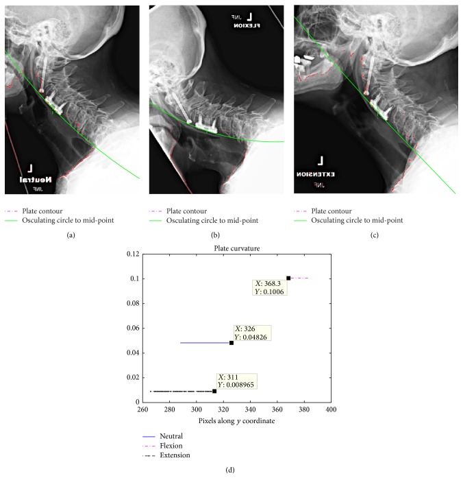
We present a new analysis tool for cervical flexion-extension radiographs based on machine vision and computerized image processing. The method is based on semiautomatic image segmentation leading to detection of common landmarks such as the spinolaminar (SL) line or contour lines of the implanted anterior cervical plates. The technique allows for visualization of the local curvature of these landmarks during flexion-extension experiments. In addition to changes in the curvature of the SL line, it has been found that the cervical plates also deform during flexion-extension examination. While extension radiographs reveal larger curvature changes in the SL line, flexion radiographs on the other hand tend to generate larger curvature changes in the implanted cervical plates. Furthermore, while some lordosis is always present in the cervical plates by design, it actually decreases during extension and increases during flexion. Possible causes of this unexpected finding are also discussed. The described analysis may lead to a more precise interpretation of flexion-extension radiographs, allowing diagnosis of spinal instability and/or pseudoarthrosis in already seemingly fused spines.
Figures
Similar articles
-
Use of flexion and extension radiographs of the cervical spine to rule out acute instability in patients with negative computed tomography scans.J Orthop Trauma. 2011 Jan;25(1):51-6. doi: 10.1097/BOT.0b013e3181dc54bf. J Orthop Trauma. 2011. PMID: 21085024
-
Sagittal alignment of cervical flexion and extension: lateral radiographic analysis.Spine (Phila Pa 1976). 2002 Aug 1;27(15):E348-55. doi: 10.1097/00007632-200208010-00014. Spine (Phila Pa 1976). 2002. PMID: 12163735
-
Utility of flexion-extension radiographs in evaluating the degenerative cervical spine.Spine (Phila Pa 1976). 2007 Apr 20;32(9):975-9. doi: 10.1097/01.brs.0000261409.45251.a2. Spine (Phila Pa 1976). 2007. PMID: 17450072
-
Cervical flexion and extension radiographs in acutely injured patients.Clin Orthop Relat Res. 1999 Aug;(365):111-6. doi: 10.1097/00003086-199908000-00015. Clin Orthop Relat Res. 1999. PMID: 10627694
-
Cervical instability in cervical spondylosis patients : Significance of the radiographic index method for evaluation.Orthopade. 2018 Dec;47(12):977-985. doi: 10.1007/s00132-018-3635-3. Orthopade. 2018. PMID: 30255359 Free PMC article.
Cited by
-
Computer Vision Based Automatic Extraction and Thickness Measurement of Deep Cervical Flexor from Ultrasonic Images.Comput Math Methods Med. 2016;2016:5892051. doi: 10.1155/2016/5892051. Epub 2016 Feb 1. Comput Math Methods Med. 2016. PMID: 26949411 Free PMC article.
-
Intelligent Automatic Segmentation of Wrist Ganglion Cysts Using DBSCAN and Fuzzy C-Means.Diagnostics (Basel). 2021 Dec 10;11(12):2329. doi: 10.3390/diagnostics11122329. Diagnostics (Basel). 2021. PMID: 34943564 Free PMC article.
References
-
- Omeis I., DeMattia J. A., Hillard V. H., Murali R., Das K. History of instrumentation for stabilization of the subaxial cervical spine. Neurosurgical Focus. 2004;16(1, article E10) - PubMed
-
- King J. T., Jr., Abbed K. M., Gould G. C., Benzel E. C., Ghogawala Z. Cervical spine reoperation rates and hospital resource utilization after initial surgery for degenerative cervical spine disease in 12 338 patients in Washington state. Neurosurgery. 2009;65(6):1011–1022. doi: 10.1227/01.NEU.0000360347.10596.BD. - DOI - PubMed
-
- Raizman N. M., O'brien J. R., Poehling-Monaghan K. L., Yu W. D. Pseudarthrosis of the spine. Journal of the American Academy of Orthopaedic Surgeons. 2009;17(8):494–503. - PubMed
LinkOut - more resources
Full Text Sources
Other Literature Sources