

Disrupted balance of T cells under natalizumab treatment in multiple sclerosis
- PMID: 27006971
- PMCID: PMC4784802
- DOI: 10.1212/NXI.0000000000000210
Disrupted balance of T cells under natalizumab treatment in multiple sclerosis
Abstract
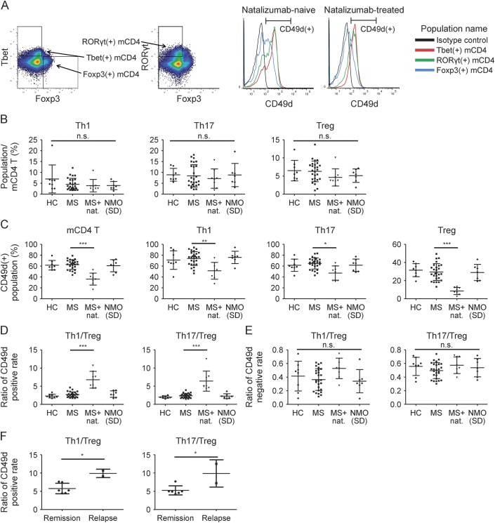
Objective: To compare effects of natalizumab on inflammatory and regulatory T cells with regard to expression of α4-integrin (CD49d).
Methods: Twenty-seven natalizumab-naive and 8 natalizumab-treated patients with multiple sclerosis (MS), 7 patients with neuromyelitis optica (NMO) or NMO spectrum disorder, and 8 healthy controls were included. The positive rate of CD49d was analyzed and compared among T helper 1 (Th1), T helper 17 (Th17), and regulatory T (Treg) cells (CD49d+Th1, CD49d+Th17, and CD49d+Treg, respectively).
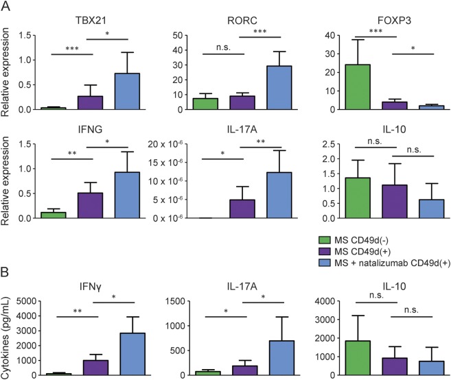
Results: Natalizumab treatment increased CD49d ratios, CD49d+Th1/CD49d+Treg, and CD49d+Th17/CD49d+Treg. This indicates larger reduction of the CD49d+ population in Treg cells than in Th1 or Th17 cells. The CD49d ratios of 2 patients who experienced exacerbation during natalizumab treatment were remarkably higher than those of the other natalizumab-treated patients. Natalizumab treatment increased the expression of TBX21, RORC, interferon (IFN)-γ, and interleukin (IL)-17A, and decreased the expression of FOXP3 in CD49d+ memory CD4 T cells. Natalizumab treatment also increased the amount of IFN-γ and IL-17A secreted by CD49d+ memory CD4 T cells.
Conclusions: The reduction rate of the CD49d+ population in Treg cells was larger than that in Th1 or Th17 cells. Although the large reduction in CD49d+ population is beneficial for MS, the proinflammatory state of residual CD49d+ cells might, in part, explain the presence of disease activity under natalizumab treatment.
Figures
References
-
- Stuve O, Gold R, Chan A, Mix E, Zettl U, Kieseier BC. alpha4-Integrin antagonism with natalizumab: effects and adverse effects. J Neurol 2008;255(suppl 6):58–65. - PubMed
-
- Yednock TA, Cannon C, Fritz LC, Sanchez-Madrid F, Steinman L, Karin N. Prevention of experimental autoimmune encephalomyelitis by antibodies against alpha 4 beta 1 integrin. Nature 1992;356:63–66. - PubMed
-
- Havrdova E, Galetta S, Hutchinson M, et al. Effect of natalizumab on clinical and radiological disease activity in multiple sclerosis: a retrospective analysis of the Natalizumab Safety and Efficacy in Relapsing-Remitting Multiple Sclerosis (AFFIRM) study. Lancet Neurol 2009;8:254–260. - PubMed
-
- Prosperini L, Gianni C, Barletta V, et al. Predictors of freedom from disease activity in natalizumab treated-patients with multiple sclerosis. J Neurol Sci 2012;323:104–112. - PubMed
LinkOut - more resources
Full Text Sources
Other Literature Sources
Molecular Biology Databases
Research Materials