. 2016:2016:9561968.
doi: 10.1155/2016/9561968. Epub 2016 Feb 23.

Tamoxifen Promotes Axonal Preservation and Gait Locomotion Recovery after Spinal Cord Injury in Cats

Affiliations

Tamoxifen Promotes Axonal Preservation and Gait Locomotion Recovery after Spinal Cord Injury in Cats

Braniff de la Torre Valdovinos et al. J Vet Med. 2016.

Abstract

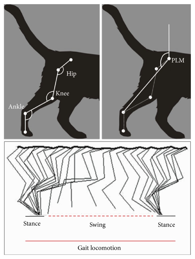
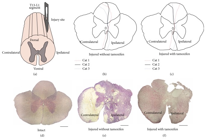
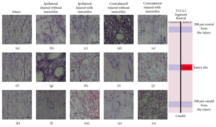
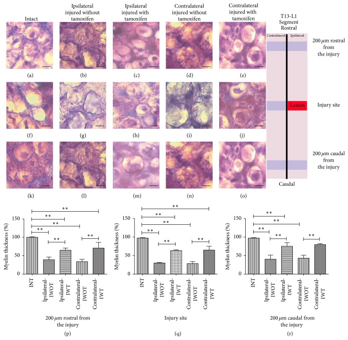
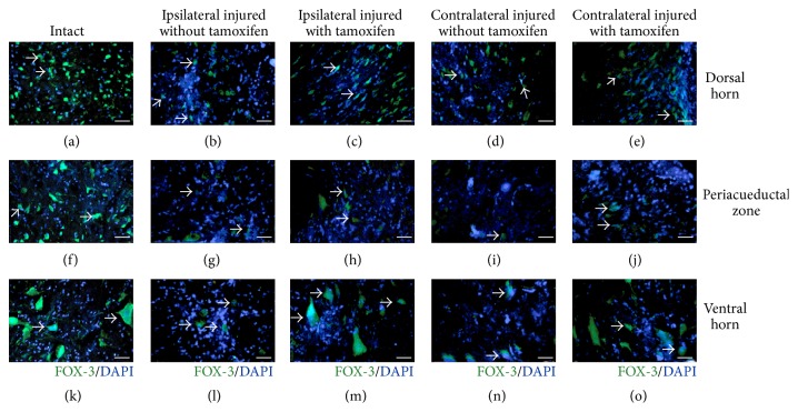
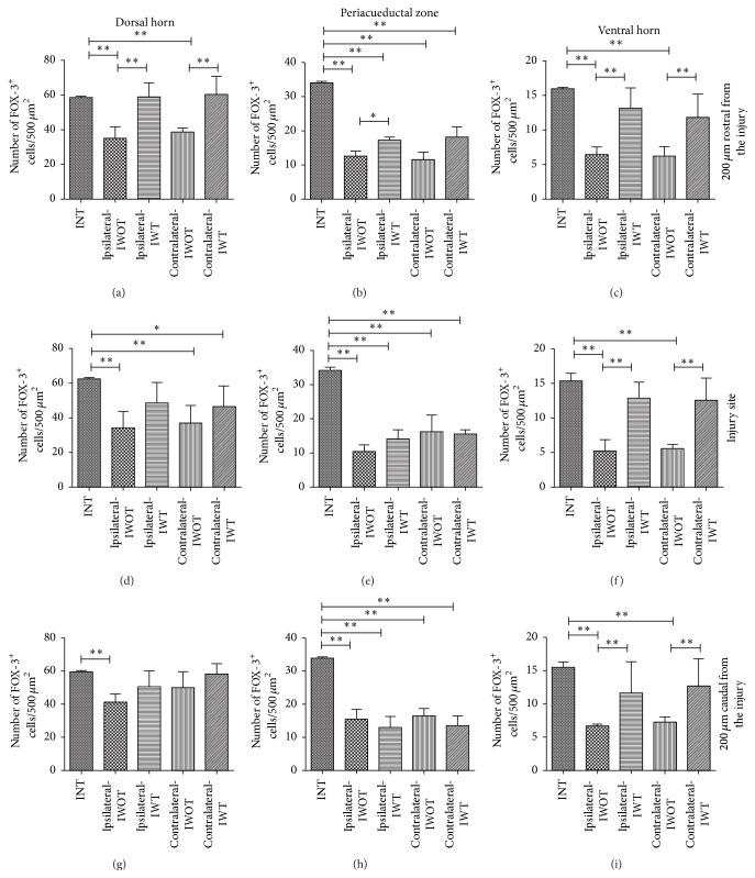
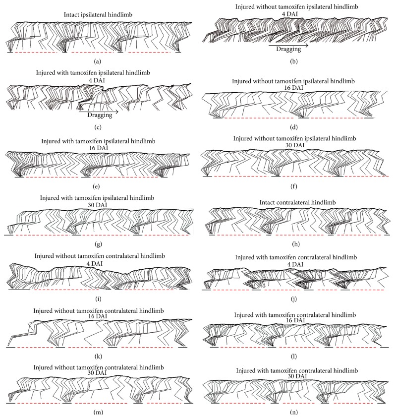
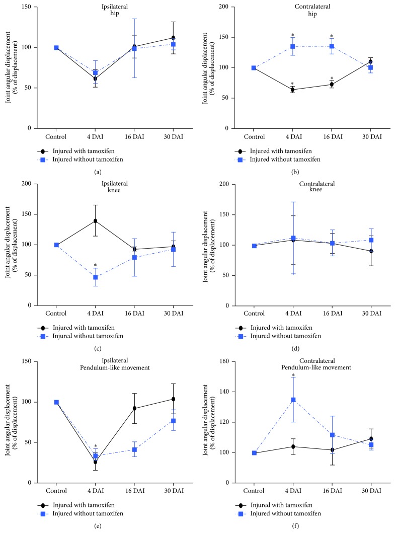
We performed experiments in cats with a spinal cord penetrating hemisection at T13-L1 level, with and without tamoxifen treatment. The results showed that the numbers of the ipsilateral and contralateral ventral horn neurons were reduced to less than half in the nontreated animals compared with the treated ones. Also, axons myelin sheet was preserved to almost normal values in treated cats. On the contrary, in the untreated animals, their myelin sheet was reduced to 28% at 30 days after injury (DAI), in both the ipsilateral and contralateral regions of the spinal cord. Additionally, we made hindlimb kinematics experiments to study the effects of tamoxifen on cat locomotion after the injury: at 4, 16, and 30 DAI. We observed that the ipsilateral hindlimb angular displacement (AD) of the pendulum-like movements (PLM) during gait locomotion was recovered to almost normal values in treated cats. Contralateral PLM acquired similar values to those obtained in intact cats. At 4 DAI, untreated animals showed a compensatory increment of PLM occurring in the contralateral hindlimb, which was partially recovered at 30 DAI. Our findings indicate that tamoxifen exerts a neuroprotective effect and preserves or produces myelinated axons, which could benefit the locomotion recovery in injured cats.


Figures

Figure 1
Figure 1
Diagram illustrating the experimental arrangement. Figure shows a schematic representation of the hip, knee, ankle, and pendulum like-movement (PLM) angles as well as the swing and stance step phases.
Figure 2
Figure 2
Representative T13-L1 microscopy images stained with H&E in coronal cuts (20 μm thick) and schematic illustrations showing the lesion extension size. (a) Schematic diagram T13-L1 segment; (b) schematic diagram illustrating the lesion extension size for each one of the three cats injured without tamoxifen group; black continuous line represents cat 1, red circular dotted line cat 2, and blue oval dotted line cat 3; (c) schematic diagram showing lesions extensions in each one of the cats in the injured with tamoxifen group, lines as indicated. (d) Spinal cord coronal cut from an intact cat. (e) Spinal cord coronal cut from an injured cat without tamoxifen. (f) Spinal cord cut from an injured cat with tamoxifen; scale bar: 500 μm.
Figure 3
Figure 3
Illustrates ventral T13-L1 axons using H&E staining. ((a)–(o)) exhibits ventral axons of the white matter of the intact, injured treated with tamoxifen, and injured without tamoxifen cats in the ipsilateral and contralateral sides. ((a), (f), and (k)) Intact cat, ((b), (g), and (l)) ipsilateral side coronal cuts in an injured cat without tamoxifen, ((c), (h), and (m)) ipsilateral side coronal cuts from an injured cat treated with tamoxifen, ((d), (i), and (n)) contralateral coronal cuts obtained in an injured cat without tamoxifen, and ((e), (j), and (o)) contralateral coronal cut obtained from an injured with tamoxifen cat.
Figure 4
Figure 4
Myelin sheet of T13-L1 ventral axons is illustrated using Toluidine Blue staining. ((a), (f), and (k)) Axons in coronal cuts from an intact cat, ((b), (g), and (l)) ipsilateral axons from an injured cat without tamoxifen, ((c), (h), and (m)) ipsilateral axons from an injured cat with tamoxifen, ((d), (i), and (n)) contralateral axons in a cat without tamoxifen, and ((e), (j), and (o)) contralateral axons in a cat with tamoxifen. Graphs: ordinates exhibit the myelin thickness percentage from ventral zone axons; abscise different studied groups. (p) Myelin axon thickness valued at 200 μm rostral from the injury, (q) axon myelin thickness valued in the injury site, (r) axon myelin thickness valued 200 μm caudal from the injury situ, ∗∗ p < 0.001; scale bar: 5 μm. Schematic diagram illustrates in site the rostral and caudal zones to value myelin thickness. The black horizontal line over the bars indicates a statistical significant difference between referred groups.
Figure 5
Figure 5
Microscopy images of FOX-3/DAPI positive cells in the dorsal horn, periaqueductal zone, and ventral horn, at T13-L1 spinal cord injury site. Dorsal horn neurons: ((b) and (c)) ipsilateral side, ((d) and (e)) contralateral side: (a) intact cat, (b) injured untreated cat, (c) injured treated cat, (d) injured untreated cat, and (e) injured treated cat. Periaqueductal Zone Neurons: ((g) and (h)) ipsilateral side and ((i) and (j)) contralateral side. (f) Intact cat, (g) injured cat without tamoxifen, (h) injured cat with tamoxifen, (i) injured cat without tamoxifen, and (j) injured cat with tamoxifen. Neurons in the ventral horn: ((l) and (m)) ipsilateral side and ((n) and (o)) contralateral side. (k) Intact cat, (l) injured cat without tamoxifen, (m) injured cat with tamoxifen, (n) injured cat without tamoxifen, and (o) injured cat with tamoxifen, scale bar 150 μm; FOX-3 neurons in green, cell nuclei in blue.
Figure 6
Figure 6
Number of counted neurons at the studied sites, graphs ordinates exhibited the number of FOX-3/DAPI positive cells and abscise different groups: intact, injured with tamoxifen, and injured without tamoxifen cats. Number of neurons 200 μm rostral from the injury site: (a) dorsal horn, (b) periaqueductal zone, and (c) ventral horn. Number of neurons in the injury site: (d) dorsal horn, (e) periaqueductal zone, and (f) ventral horn. Number of neurons counted 200 μm caudal from the injury: (g) dorsal horn, (h) periaqueductal zone, and (i) ventral horn; ∗∗ p < 0.001, p < 0.05. Schematic diagram illustrates rostral, injury site and caudal zones. The black horizontal line over bars indicates a statistically significant difference between referred groups.
Figure 7
Figure 7
Stick figures illustrating both cat hindlimb during overground locomotion in intact and in spinal hemisected cats treated with tamoxifen. ((a)–(n)) Stick figures illustrating gait locomotion in intact cat and after 4, 16, and 30 DAI: in treated and untreated cats. (a) Ipsilateral hindlimb during locomotion in a intact cat. (b) Ipsilateral hindlimb during locomotion in an untreated cat at 4 DAI; arrow shows dragging during forward displacement. (c) Ipsilateral hindlimb locomotion in treated cat, at 4 DAI. (d) Ipsilateral hindlimb locomotion in an untreated cat, at 16 DP. (e) Ipsilateral hindlimb, in treated cat, at 16 DAI. (f) Ipsilateral hindlimb in an untreated cat at 30 DAI. (g) Ipsilateral hindlimb locomotion in treated cat, at 30 DAI. (h) Contralateral hindlimb locomotion in an intact cat. (i) Contralateral hindlimb locomotion in an untreated cat, at 4 DAI. (j) Contralateral hindlimb in treated cat, at 4 DAI. (k) Contralateral hindlimb locomotion in an untreated cat, at 16 DAI. (l) Contralateral hindlimb in treated cat, at 16 DAI. (m) Contralateral hindlimb locomotion in a untreated cat, at 30 DAI. (n) Contralateral hindlimb locomotion in treated cat, at 30 DAI.
Figure 8
Figure 8
Graphs exhibit hip, knee joints, and the hindlimb pendulum-like movement angular displacement during stride. Movement changes in percent at different times (4, 16, and 30 DAI) comparing the respective angles and pendulum-like movement previous to the injury (control). ((a) and (b)) Hip angular displacement, ((c) and (d)) knee angular displacement, and ((e) and (f)) pendulum-like movement angular displacement; p < 0.05.

Similar articles

Cited by

References

    1. Jacobs R. R., Asher M. A., Snider R. K. Thoracolumbar spinal injuries. A comparative study of recumbent and operative treatment in 100 patients. Spine. 1980;5(5):463–477. doi: 10.1097/00007632-198009000-00012. - DOI - PubMed
    1. Guptarak J., Wiktorowicz J. E., Sadygov R. G., et al. The cancer drug tamoxifen: a potential therapeutic treatment for spinal cord injury. Journal of Neurotrauma. 2014;31(3):268–283. doi: 10.1089/neu.2013.3108. - DOI - PMC - PubMed
    1. Tester N. J., Howland D. R. Chondroitinase ABC improves basic and skilled locomotion in spinal cord injured cats. Experimental Neurology. 2008;209(2):483–496. doi: 10.1016/j.expneurol.2007.07.019. - DOI - PMC - PubMed
    1. Rossignol S., Giroux N., Chau C., Marcoux J., Brustein E., Reader T. A. Pharmacological aids to locomotor training after spinal injury in the cat. Journal of Physiology. 2001;533(1):65–74. doi: 10.1111/j.1469-7793.2001.0065b.x. - DOI - PMC - PubMed
    1. Pertici V., Pin-Barre C., Felix M.-S., Laurin J., Brisswalter J., Decherchi P. A new method to assess weight-bearing distribution after central nervous system lesions in rats. Behavioural Brain Research. 2014;259:78–84. doi: 10.1016/j.bbr.2013.10.043. - DOI - PubMed