. 2016 Mar 23;11(3):e0149106.
doi: 10.1371/journal.pone.0149106. eCollection 2016.

Effects of Plectin Depletion on Keratin Network Dynamics and Organization

Affiliations

Effects of Plectin Depletion on Keratin Network Dynamics and Organization

Marcin Moch et al. PLoS One. .

Abstract

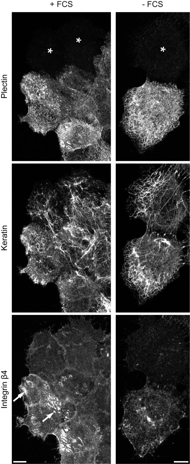
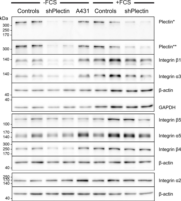
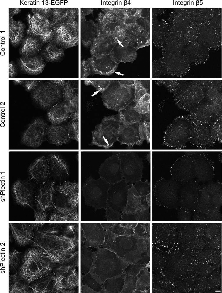
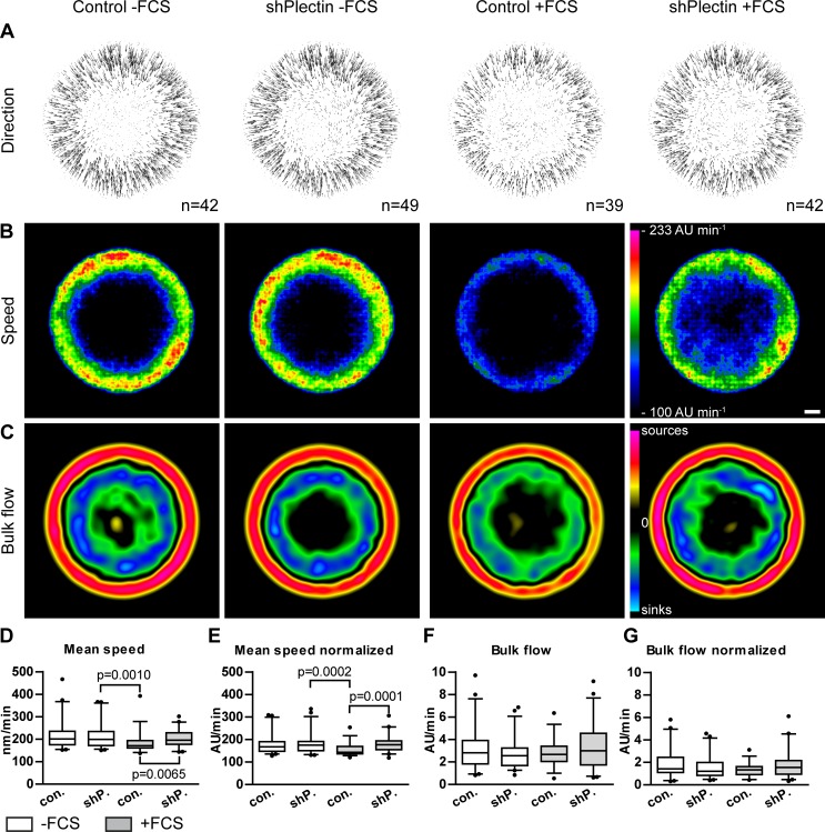
The keratin intermediate filament cytoskeleton protects epithelial cells against various types of stress and is involved in fundamental cellular processes such as signaling, differentiation and organelle trafficking. These functions rely on the cell type-specific arrangement and plasticity of the keratin system. It has been suggested that these properties are regulated by a complex cycle of assembly and disassembly. The exact mechanisms responsible for the underlying molecular processes, however, have not been clarified. Accumulating evidence implicates the cytolinker plectin in various aspects of the keratin cycle, i.e., by acting as a stabilizing anchor at hemidesmosomal adhesion sites and the nucleus, by affecting keratin bundling and branching and by linkage of keratins to actin filament and microtubule dynamics. In the present study we tested these hypotheses. To this end, plectin was downregulated by shRNA in vulvar carcinoma-derived A431 cells. As expected, integrin β4- and BPAG-1-positive hemidesmosomal structures were strongly reduced and cytosolic actin stress fibers were increased. In addition, integrins α3 and β1 were reduced. The experiments furthermore showed that loss of plectin led to a reduction in keratin filament branch length but did not alter overall mechanical properties as assessed by indentation analyses using atomic force microscopy and by displacement analyses of cytoplasmic superparamagnetic beads using magnetic tweezers. An increase in keratin movement was observed in plectin-depleted cells as was the case in control cells lacking hemidesmosome-like structures. Yet, keratin turnover was not significantly affected. We conclude that plectin alone is not needed for keratin assembly and disassembly and that other mechanisms exist to guarantee proper keratin cycling under steady state conditions in cultured single cells.


Conflict of interest statement

Competing Interests: The authors have declared that no competing interests exist.

Figures

Fig 1
Fig 1. Plectin knock down prevents formation of integrin β4 clusters and slightly affects overall keratin network morphology in A431 cells.
Vulvar carcinoma derived A431 cells were plated on laminin 332-rich matrix 48 h prior to methanol/acetone fixation. The images show triple immunofluorescence staining of plectin (guinea pig antibodies), integrin β4, and pan-keratin. They demonstrate the efficient down regulation of plectin by shRNA in cells marked with an asterisk in comparison to non-transfected neighboring cells. They also show that hemidesmosomal integrin β4 clusters (arrows) are formed in the presence of FCS in contrast to plectin-free cells and serum-starved cells (+FCS vs. -FCS). Note the less dense and more compact keratin filament networks in plectin-depleted cells. All images are maximum intensity projections from confocal sectioning of complete cells. Bars, 10 μm.
Fig 2
Fig 2. Immunoblots detect efficient reduction of plectin and complex alterations of integrin expression in A431 clones stably transfected with plectin shRNAs.
Complete cell lysates were obtained from AK13-1-derived subclones that were either stably transfected with scramble shRNA (controls) or plectin shRNA (shPlectin). For comparison, cell lysates from non-transfected A431 cells were prepared. The cells had been cultivated on laminin 332-rich matrices for 48 h either without (left) or with FCS (right). Proteins were separated by 8% SDS-PAGE. Note the efficient down regulation of plectin by 80–90% in both plectin shRNA clones as detected by two different antibodies (*guinea pig, **mouse). In addition, integrins β1 and α3 are also noticeably reduced in plectin-deficient cells. Integrin β4 is only reduced in serum-starved cells upon plectin depletion. The changes in expression levels of other integrins are apparently not linked to plectin expression. Note also, that integrin expression is increased in the presence of serum.
Fig 3
Fig 3. Immunofluorescence microscopy shows efficient down regulation of plectin in stably transfected A431 clones producing plectin shRNA.
The fluorescence images (maximum intensity projections of complete cells) were recorded in AK13-1 subclones stably expressing HK13-EGFP and either scramble control shRNAs or plectin shRNAs. The cells were grown for 48 h on laminin 332-rich matrices in the absence of FCS prior to methanol/acetone fixation. Note the very low level of plectin immunoreactivity (guinea pig antibodies) in the clones expressing plectin shRNAs in contrast to the strong plectin signal in the scramble controls presenting filamentous fluorescence co-localizing in part with the endogenous keratin filaments. The HK13-EGFPnetwork is less dense and more compact in the plectin-deficient clones. Bar, 10 μm.
Fig 4
Fig 4. Integrin β4 clusters are almost absent in plectin-deficient cells while integrin β5 clusters persist.
Immunocytochemistry of A431 clones stably expressing HK13-EGFP and either scramble control shRNA or plectin shRNA. Cells were cultivated for 48 h on laminin 332-rich matrix in the presence of FCS. The confocal images show only the bottom planes. The control cells present hemidesmosome-like integrin β4-positive clusters (arrows) whereas the plectin-deficient clones lack them and show only a rather weak and diffuse integrin β4 signal. Integrin β5 staining indicates the continued presence of focal adhesions in all clones. Keratin networks marked with HK13-EGFP are found to be barely perturbed in plectin-deficient clones. Bar, 10 μm.
Fig 5
Fig 5. Plectin-deficiency results in decreased keratin network branch length but does not alter overall cellular stiffness and cytoplasmic viscoelasticity.
A431 cells (wild type) and AK13-1 subclones stably expressing HK13-EGFP and either scramble control shRNAs or plectin shRNAs were grown for 48–57 h on laminin 332-rich matrices. A Histogram of branch length-measurements. Confocal fluorescence images of the bottom plane of isolated single cells were recorded to measure the HK13-EGFP filament length between branch points by automated image analysis. Results from either scramble control shRNA clones 1 and 2 or plectin shRNA clones 1 and 2 were combined. The plectin-deficient cells have significantly increased branch length in the absence of FCS which is even more apparent in the presence of FCS. The whiskers are min to max and statistical differences were determined by two-tailed Mann-Whitney test. B 3D confocal microscopy (maximum intensity projections are shown) of live cells after 48 h growth in the presence of 10% FCS. Note that mesh size and bundling of the keratin filament network is increased in the plectin-deficient cell. C Histogram depicting apparent stiffness A1.8 of single cells that was determined from indentation experiments that were performed above the nucleus in plectin shRNA clone 1 and scramble control clone 1. Error bars are standard deviation. D Graph showing the average forces needed to reach different indentation depths above the nucleus (same cells as in C). E Creep compliance measurements of magnetic tweezers experiments. The rescaled peaks [nshPlectin = 117 (20 cells), ncontrol = 107 (19 cells), nwild type = 67 (12 cells)] were fitted to equation 1 and averaged for each phenotype. Error bars are standard deviation. Note that no significant differences in viscoelastic properties of the cytoplasm were observed.
Fig 6
Fig 6. Keratin filament dynamics are affected by plectin-deficiency in the presence of serum but not in serum-starved cells.
AK13-1 subclones stably expressing HK13-EGFP and either scramble control shRNAs (clones 1 and 2) or plectin shRNAs (clones 1 and 2) were cultivated for 48–57 h on laminin 332-rich matrices. HK13-EGFP dynamics were recorded by confocal microscopy in the bottom plane of isolated single cells at 30 s intervals for 10 minutes. For analysis, data from control clones and plectin shRNA clones were merged together in each case. A-C show averaged results from cells that were converted to standardized circular cells. The direction and speed of movement is represented by vector fields in the top panel. Color-coded heat maps of speed of keratin are depicted in the middle panel in arbitrary units (AU) per minute. The lower panel presents derived turnover maps revealing zones of keratin assembly (sources) and disassembly (sinks). Scale bar is 5000 AU. In D-G corresponding box plots for keratin mean speed and total bulk flow are shown as determined for non-normalized networks and normalized networks. The keratin movement in plectin-depleted cells is significantly increased in comparison to control cells cultivated in the presence of FCS, which induces the formation of hemidesmosome like structures. Otherwise, plectin has no influence on keratin movement in cells cultivated without FCS. Note that the detected differences in keratin turnover were too small to be significant. Statistical analysis was performed with two-tailed Mann-Whitney test and whiskers are 5–95%.

Similar articles

Cited by

References

    1. Loschke F, Seltmann K, Bouameur JE, Magin TM. Regulation of keratin network organization. Curr Opin Cell Biol. 2015;32C:56–64. 10.1016/j.ceb.2014.12.006 . - DOI - PubMed
    1. Pan X, Hobbs RP, Coulombe PA. The expanding significance of keratin intermediate filaments in normal and diseased epithelia. Curr Opin Cell Biol. 2013;25(1):47–56. Epub 2012/12/29. 10.1016/j.ceb.2012.10.018 - DOI - PMC - PubMed
    1. Herrmann H, Strelkov SV, Burkhard P, Aebi U. Intermediate filaments: primary determinants of cell architecture and plasticity. J Clin Invest. 2009;119(7):1772–83. Epub 2009/07/10. 10.1172/JCI38214 - DOI - PMC - PubMed
    1. Iwatsuki H, Suda M. Seven kinds of intermediate filament networks in the cytoplasm of polarized cells: structure and function. Acta histochemica et cytochemica. 2010;43(2):19–31. 10.1267/ahc.10009 - DOI - PMC - PubMed
    1. Toivola DM, Strnad P, Habtezion A, Omary MB. Intermediate filaments take the heat as stress proteins. Trends Cell Biol. 2010;20(2):79–91. 10.1016/j.tcb.2009.11.004 - DOI - PMC - PubMed

Publication types

MeSH terms