Review
. 2016 Aug;73(15):2871-96.
doi: 10.1007/s00018-016-2180-7. Epub 2016 Mar 23.

Using mitochondrial sirtuins as drug targets: disease implications and available compounds

Affiliations
Review

Using mitochondrial sirtuins as drug targets: disease implications and available compounds

Melanie Gertz et al. Cell Mol Life Sci. 2016 Aug.

Abstract

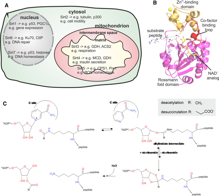
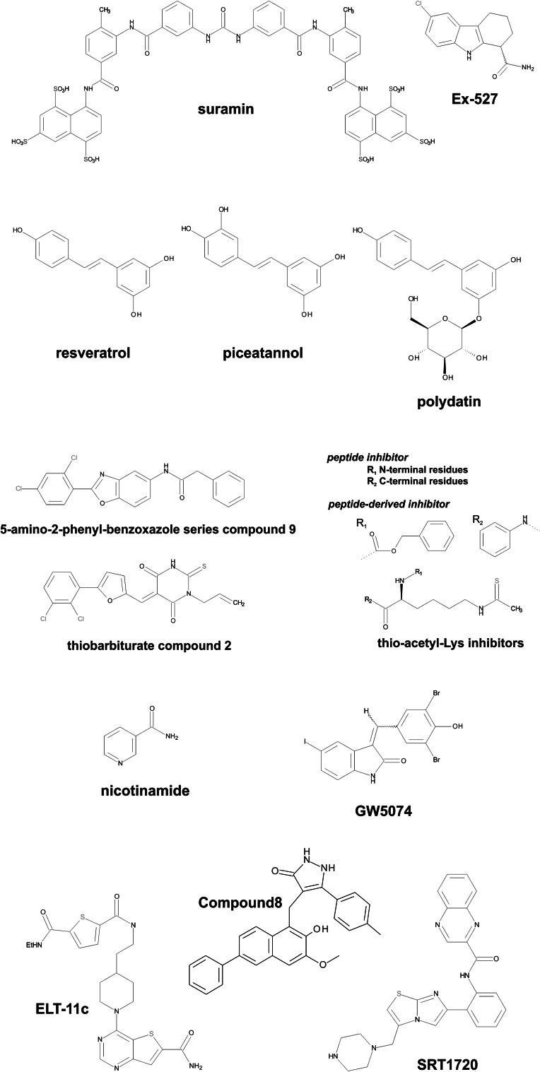
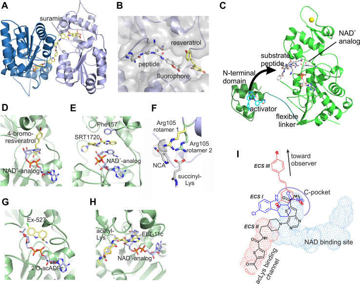
Sirtuins are an evolutionary conserved family of NAD(+)-dependent protein lysine deacylases. Mammals have seven Sirtuin isoforms, Sirt1-7. They contribute to regulation of metabolism, stress responses, and aging processes, and are considered therapeutic targets for metabolic and aging-related diseases. While initial studies were focused on Sirt1 and 2, recent progress on the mitochondrial Sirtuins Sirt3, 4, and 5 has stimulated research and drug development for these isoforms. Here we review the roles of Sirtuins in regulating mitochondrial functions, with a focus on the mitochondrially located isoforms, and on their contributions to disease pathologies. We further summarize the compounds available for modulating the activity of these Sirtuins, again with a focus on mitochondrial isoforms, and we describe recent results important for the further improvement of compounds. This overview illustrates the potential of mitochondrial Sirtuins as drug targets and summarizes the status, progress, and challenges in developing small molecule compounds modulating their activity.

Keywords: Activator; Deacylase; Drug development; Inhibitor; Metabolic regulation; Sirt3; Sirt4; Sirt5.


Conflict of interest statement

The authors declare that they have no conflict of interest.

Figures

Fig. 1
Fig. 1
Localization and functions of mammalian Sirtuin isoforms and architecture and mechanism of their catalytic domains. a Schematic view on the main subcellular localization of mammalian Sirtuin isoforms. Examples for major functions and substrates are indicated. b Cartoon presentation of the structure of human Sirt3 with bound AceCS2 substrate peptide and NAD+ analog (ligands as sticks; PDB ID 4FVT [232]). The large Rossmann fold domain, the small Zn2+ binding domain, and the cofactor binding loop are indicated; the Zn2+ ion is shown as a sphere. c Reaction mechanism of Sirtuin-catalyzed, NAD+-dependent deacylations
Fig. 2
Fig. 2
Chemical structures of compounds with significant effects on the activities of mitochondrial Sirtuins
Fig. 3
Fig. 3
Structural analysis of Sirt3 and Sirt5 modulation by small molecule drugs. a Crystal structure of Sirt5 in complex with suramin (PDB ID 2NYR; [222]), which inhibits Sirtuins non-specifically by blocking the active site and artificially crosslinks two monomers. b Crystal structure of Sirt5 in complex with resveratrol and fluorophore-modified peptide (PDB ID 4HDA; [201]) showing the direct contact between substrate and activator. c Crystal structure of Sirt1 with substrate peptide and activator (grey sticks) bound to the active site of the catalytic core (green), and with an activator (cyan sticks) bound to the Sirt1-specific N-terminal domain (dark green). The N-terminal domain is speculated to swing, via the flexible linker, toward the active site (arrow) and to cover it lid-like with the exposed surface of the activator, resulting in a direct activator/substrate contact. d Crystal structure of Sirt3 in complex with 4-bromo-resveratrol (PDB ID 4C7B [203]). The NAD+-analog carba-NAD+ was modeled through an overlay with a Sirt3/ACS2-peptide/carba-NAD+ structure (PDB ID 4FVT; [239]) to illustrate that the inhibitor blocks the binding pocket for the NAD+ nicotinamide group. e Crystal structure of Sirt3 in complex with SRT1720 (PDB ID 4BN5; [187]) and the NAD+-analog carba-NAD+ showing the extensive interaction between these two uncompetitive ligands. f Model for the molecular basis of Sirt5’s differential nicotinamide sensitivity. The structure of Sirt5 in complex with succinylated peptide and NAD+ (PDB ID 3RIY; [151]) shows an interaction between Arg105 (green, rotamer 2) and the substrate succinylate. In absence of this interaction, for example when an acetylated substrate is bound, a preferred conformation of Arg105 (yellow, rotamer 1) would clash with nicotinamide (NCA; modelled into its known binding pocket). g Crystal structure of Sirt3 in complex with Ex-527 and the co-product 2′-O-acetyl-ADP-ribose (PDB ID 4BVH; [179]), which increases inhibitor affinity apparently by stabilizing a closed active site conformation. h Crystal structure of Sirt3 in complex with ELT-11c (PDB ID 4JSR; [202]). Acetyl-Lys and the NAD+-analog carba-NAD+ were modeled in through an overlay with the complex structure Sirt3/ACS2-peptide/carba-NAD+ (PDB ID 4FVT; [239]) to illustrate that the inhibitor occupies the acetyl-Lys channel und the binding region for the NAD+ nicotinamide riboside. i Scheme for the binding modes of Sirtuin inhibitors exploiting the C site

References

    1. Choudhary C, Kumar C, Gnad F, Nielsen ML, Rehman M, Walther TC, Olsen JV, Mann M. Lysine acetylation targets protein complexes and co-regulates major cellular functions. Science. 2009;325:834–840. doi: 10.1126/science.1175371. - DOI - PubMed
    1. Norvell A, McMahon SB. Cell biology. Rise of the rival. Science. 2010;327:964–965. doi: 10.1126/science.1187159. - DOI - PubMed
    1. Rauh D, Fischer F, Gertz M, Lakshminarasimhan M, Bergbrede T, Aladini F, Kambach C, Becker CFW, Zerweck J, Schutkowski M, Steegborn C. An acetylome peptide microarray reveals specificities and deacetylation substrates for all human sirtuin isoforms. Nat Commun. 2013;4:2327. doi: 10.1038/ncomms3327. - DOI - PubMed
    1. Rardin MJ, Newman JC, Held JM, Cusack MP, Sorensen DJ, Li B, Schilling B, Mooney SD, Kahn CR, Verdin E, Gibson BW. Label-free quantitative proteomics of the lysine acetylome in mitochondria identifies substrates of SIRT3 in metabolic pathways. Proc Natl Acad Sci USA. 2013;110:6601–6606. doi: 10.1073/pnas.1302961110. - DOI - PMC - PubMed
    1. Still AJ, Floyd BJ, Hebert AS, Bingman CA, Carson JJ, Gunderson DR, Dolan BK, Grimsrud PA, Dittenhafer-Reed KE, Stapleton DS, Keller MP, Westphall MS, Denu JM, Attie AD, Coon JJ, Pagliarini DJ. Quantification of mitochondrial acetylation dynamics highlights prominent sites of metabolic regulation. J Biol Chem. 2013;288:26209–26219. doi: 10.1074/jbc.M113.483396. - DOI - PMC - PubMed

Publication types

MeSH terms

LinkOut - more resources