. 2016 Nov;9(6):1571-1583.
doi: 10.1038/mi.2016.22. Epub 2016 Mar 23.

Medroxyprogesterone acetate and levonorgestrel increase genital mucosal permeability and enhance susceptibility to genital herpes simplex virus type 2 infection

Affiliations

Medroxyprogesterone acetate and levonorgestrel increase genital mucosal permeability and enhance susceptibility to genital herpes simplex virus type 2 infection

N E Quispe Calla et al. Mucosal Immunol. 2016 Nov.

Abstract

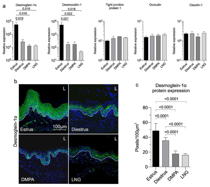
Depot-medroxyprogesterone acetate (DMPA) is a hormonal contraceptive especially popular in areas with high prevalence of HIV and other sexually transmitted infections (STI). Although observational studies identify DMPA as an important STI risk factor, mechanisms underlying this connection are undefined. Levonorgestrel (LNG) is another progestin used for hormonal contraception, but its effect on STI susceptibility is much less explored. Using a mouse model of genital herpes simplex virus type 2 (HSV-2) infection, we herein found that DMPA and LNG similarly reduced genital expression of the desmosomal cadherin desmoglein-1α (DSG1α), enhanced access of inflammatory cells to genital tissue by increasing mucosal epithelial permeability, and increased susceptibility to viral infection. Additional studies with uninfected mice revealed that DMPA-mediated increases in mucosal permeability promoted tissue inflammation by facilitating endogenous vaginal microbiota invasion. Conversely, concomitant treatment of mice with DMPA and intravaginal estrogen restored mucosal barrier function and prevented HSV-2 infection. Evaluating ectocervical biopsy tissue from women before and 1 month after initiating DMPA remarkably revealed that inflammation and barrier protection were altered by treatment identically to changes seen in progestin-treated mice. Together, our work reveals DMPA and LNG diminish the genital mucosal barrier; a first-line defense against all STI, but may offer foundation for new contraceptive strategies less compromising of barrier protection.


Conflict of interest statement

Authors have no conflict of interests to declare.

Figures

Fig 1
Fig 1. MPA and LNG increased mouse susceptibility to ivag HSV-2 infection
(a) Untreated mice in estrus and DMPA- or LNG-treated mice were ivag infected with 3 x 10⁶ pfu HSV-1q-GPF, and euthanized 1 h, 12 h, or 24 h later. Examination of excised vaginal tissue by confocal microscopy showed widespread viral dissemination in the mucosa of DMPA- and LNG-treated mice, while virus failed to extend beyond superficial mucosal surfaces of mice infected during estrus (L indicates vaginal lumen); HSV-1q-GPF (green); DAPI (blue). (b, c) In separate studies, untreated mice in estrus or diestrus; DMPA-treated, Pg-treated, DMPA and RU486-treated mice; mice treated with LNG (s.c. or ivag); or MePRDL-treated mice were infected with 104 pfu HSV-2 and observed daily for genital pathology and mortality. Consistent with fluorescent HSV data (a), Kaplan-Meier survival curve showed DMPA, LNG s.c., LNG ivag, and Pg treatments produced 100% mortality, while no mice infected in estrus, treated with DMPA and RU486, or treated with MePRDL developed morbidity or mortality (data representative of 2 independent experiments containing 5 mice per group). To compare scores for the development of genital pathology, areas under the curve were calculated for individual mice, and multiple group comparisons performed using Kruskal-Wallis test with Dunn’s post hoc test (diestrus compared to estrus, p = 0.0243; for all other groups compared to estrus, p < 0.0001). For Kaplan-Meier survival curve data, log-rank tests were used to compare cumulative survival incidence after ivag HSV-2 infection (diestrus compared to estrus, p = 0.0165; for all other groups compared to estrus, p < 0.0001).
Fig 2
Fig 2. DMPA and LNG similarly increased genital mucosal permeability in mice
(a) Vaginal mucosal thicknesses were equivalent in mice in diestrus and DMPA- or LNG-treated mice, indicating differential epithelial thinning had not cause the sizeable differences in mortality after ivag HSV-2 infection (Fig. 1b and 1c) (H&E-stained vaginal tissue sections from 4 mice were assessed per condition and 5 fields per section evaluated by light microscopy at 200X magnification). (b) Groups of mice treated as described in Fig. 1b were anesthetized and ivag administered PBS containing Lucifer yellow (LY) (457.2 Da) (green) and 70 KDa dextran-Texas Red® (DR) (red). Forty-five minutes later, mice were euthanized and vaginal tissue processed and counterstained with DAPI (blue) for confocal microscopic analysis. While neither fluorescent molecule entered vaginal tissue of mice in estrus or mice treated with DMPA and RU486 or MePRDL, LY entered the vaginal mucosa of mice in diestrus but entry was more pervasive in the vaginal mucosa of DMPA- or LNG-treated mice (representative images from 2 independent experiments with 5 animals per condition). (c) Anesthetized mice in estrus and DMPA-treated mice were ivag administered 2 x 10⁶ CFSE-labeled syngeneic splenocytes (green), and 12 hours later vaginal tissue treated as detailed in Material and Methods. Confocal microscopy analysis showed significantly deeper penetration of CFSE-labeled splenocytes into the vaginal mucosa of DMPA-treated mice vs. mice in estrus. Representative images and data quantification from 2 independent experiments with 5 animals per condition are shown (L denotes vaginal lumen). Between-group comparisons of vaginal mucosal thickness were made with the Kruskal-Wallis and Dunn’s post hoc tests, and differences in splenocyte entry into vaginal mucosa were made with the unpaired Student t test.
Fig 3
Fig 3. DMPA and LNG reduced vaginal tissue expression of the desmosomal cadherins DSG1α and DSC1
(a) Vaginal tissue was excised from uninfected, untreated mice in estrus or diestrus, and uninfected mice treated with DMPA or LNG, and processed to assess the gene expression levels of the desmosomal cadherins DSG1α and DSC1. Mice in diestrus and mice treated with MPA or LNG showed significantly lower expression DSG1α and DSC1, but not tight junction protein 1, occludin, or claudin-1 (mean ±SD) (data from 2 independent experiments with 6 mice per group). (b) Immunofluorescence staining of vaginal tissue from mice treated in same manner as (a) assessed levels of DSG1α protein expression; (representative images from 2 independent experiments with 5 animals per condition); DSG1α (green); DAPI (blue) (L denotes vaginal lumen). (c) Quantification of data displayed in (b) showed LNG and DMPA significantly reduced DSG1α protein expression compared to mice in estrus or diestrus. One-way ANOVA and Tukey’s post hoc tests provided statistical comparisons in (a) and (c).
Fig 4
Fig 4. DMPA promoted vaginal tissue inflammation by enhancing tissue invasion of endogenous vaginal microbiota
(a) Representative PNA FISH images showed dramatic increase of endogenous bacterial microbiota in the vaginal submucosa of uninfected, DMPA-treated mice (injection of Salmonella into vaginal tissue 1 h before euthanasia provided positive controls and germ-free (GF) mice provided negative controls); bacteria (green); DAPI (blue) (representative images from 2 independent experiments with 3 animals per condition). (b) qRT-PCR analysis of various proinflammatory cytokines and chemokines levels in the vaginal tissue of uninfected WT mice in estrus or diestrus, DMPA-treated mice, GF mice in estrus, or DMPA-treated GF mice showed genes related to neutrophil infiltration and inflammation were elevated in WT mice in diestrus or after DMPA treatment. However, similar responses were not seen in DMPA-treated GF mice, indicating inflammatory changes were sequelae to tissue incursion by endogenous microbiota (mean ±SD) (data displayed from 2 independent experiments with 6 mice per group). (c) Confocal microscopy images showed that the increases in vaginal mucosal permeability displayed in uninfected, DMPA-treated GF mice were not detected in untreated GF mice in estrus, implying DMPA-mediated disruption of the mucosal barrier was not sequelae to increased tissue inflammation. Representative images from 2 independent experiments with 3 animals per condition (L denotes vaginal lumen). Levels of the proinflammatory cytokines and cytokines in (b) were compared using one-way ANOVA and Dunnett’s post hoc test.
Fig 5
Fig 5. Exogenous estrogen (E) restored DSG1α expression and reduces susceptibility of DMPA-treated mice to ivag HSV-2 infection
(a, b) DMPA-treated mice or DMPA-treated mice concomitantly administered ivag E or systemic E2 were infected with 104 pfu HSV-2, and assessed for external genital pathology and mortality. Pathology and survival curve data demonstrated both E treatments abrogated development of morbidity and mortality seen in DMPA-treated mice (data shown representative of 2 independent experiments containing 5 mice per group). (c) Consistent with the effects of E treatment on mouse survival after ivag HSV-2 infection, DMPA-mediated decreases in vaginal tissue DSG1α expression were restored by local E and systemic E2 treatments (mean ±SD) (data from 2 independent experiments with 6 mice per group). (d) Both DSG1α protein expression and epithelial permeability were reestablished by E treatment of DMPA-treated mice (representative figures from 2 independent experiments and 3 mice per condition), for left panels: LY (green); DR (red); DAPI (blue), for right panels, DSG1α (green); DAPI (blue); (L denotes vaginal lumen). (e) DSG1α protein expression shown in (d) was quantified as defined in Materials and Methods (mean ±SD). Results from 2 independent experiments with 3 mice per group. Kruskal-Wallis test with Dunn’s post hoc tests were used for (a); one-way ANOVA and Tukey’s post hoc tests were used for comparisons in (c) and (e); and log-rank tests were used to compare cumulative survival incidence after ivag HSV-2 infection in (b).
Fig 6
Fig 6. Women initiating DMPA use displayed significant increases in ectocervical mucosal permeability
(a) Ectocervical biopsy tissues collected from women using no form of hormonal contraceptive (enrollment visit) and 1 month after initiating DMPA (follow-up visit) revealed significantly reduced DSG1 expression in tissues obtained after treatment (panel displays relative gene expression levels before and after DMPA initiation for 7 women. (b) Analogous to changes seen in uninfected mice after DMPA treatment, DMPA use correlated with increased ectocervical tissue inflammation at the follow-up visit (panel displays relative expression levels of genes related to inflammation and neutrophil migration for 7 women before and after initiating DMPA use). (c) Representative images from ectocervical biopsy tissue samples incubated with LY and DR revealed DMPA-mediated loss of mucosal barrier function that was notably similar to that seen in DMPA-treated mice (n=7) (L denotes the vaginal lumen). (d) Quantification of LY penetration depth into ectocervical mucosa of tissue biopsies from women before and after their initiation of DMPA use (n=7). (e) MPA serums levels detected in women at the enrollment and follow-up visits. f) Linear regression analysis identified a positive correlation between MPA serum concentration and LY penetration depth into ectocervical mucosal tissue (R2 value reported). Paired Student t tests were used to provide statistical comparisons in (a), (b), (d), and (e).

References

    1. Quinn TC, Overbaugh J. HIV/AIDS in women: an expanding epidemic. Science. 2010;308:1582–1583. - PubMed
    1. Morrison CS, et al. Hormonal contraceptive use, cervical ectopy, and the acquisition of cervical infections. Sex Transm Dis. 2004;31:561–567. - PubMed
    1. Baeten JM, et al. Hormonal contraceptive use, herpes simplex virus infection, and risk of HIV-1 acquisition among Kenyan women. AIDS. 2007;21:1771–1777. - PubMed
    1. Morrison CS, et al. Hormonal contraception and HIV acquisition: reanalysis using marginal structural modeling. AIDS. 2010;24:1778–1781. - PMC - PubMed
    1. Heffron R, et al. Partners in Prevention HSV/HIV Transmission Study Team. Use of hormonal contraceptives and risk of HIV-1 transmission: a prospective cohort study. Lancet Infect Dis. 2012;12:19–26. - PMC - PubMed

Publication types

MeSH terms