

The value of thyroid shielding in intraoral radiography
- PMID: 27008105
- PMCID: PMC5084701
- DOI: 10.1259/dmfr.20150407
The value of thyroid shielding in intraoral radiography
Abstract
Objectives: To evaluate the utility of the application of a thyroid shield in intraoral radiography when using rectangular collimation.
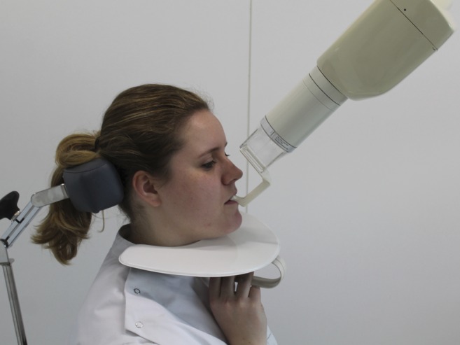
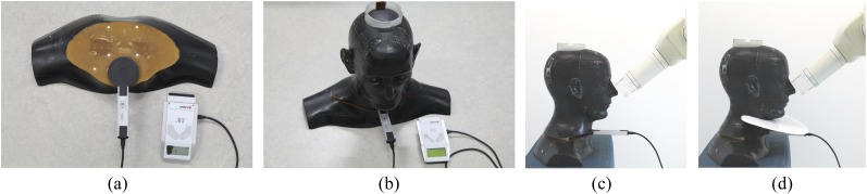
Methods: Experimental data were obtained by measuring the absorbed dose at the position of the thyroid gland in a RANDO(®) (The Phantom Laboratory, Salem, NY) male phantom with a dosemeter. Four protocols were tested: round collimation and rectangular collimation, both with and without thyroid shield. Five exposure positions were deployed: upper incisor (Isup), upper canine (Csup), upper premolar (Psup), upper molar (Msup) and posterior bitewing (BW). Exposures were made with 70 kV and 7 mA and were repeated 10 times. The exposure times were as recommended for the exposure positions for the respective collimator type by the manufacturer for digital imaging. The data were statistically analyzed with a three-way ANOVA test. Significance was set at p < 0.01.
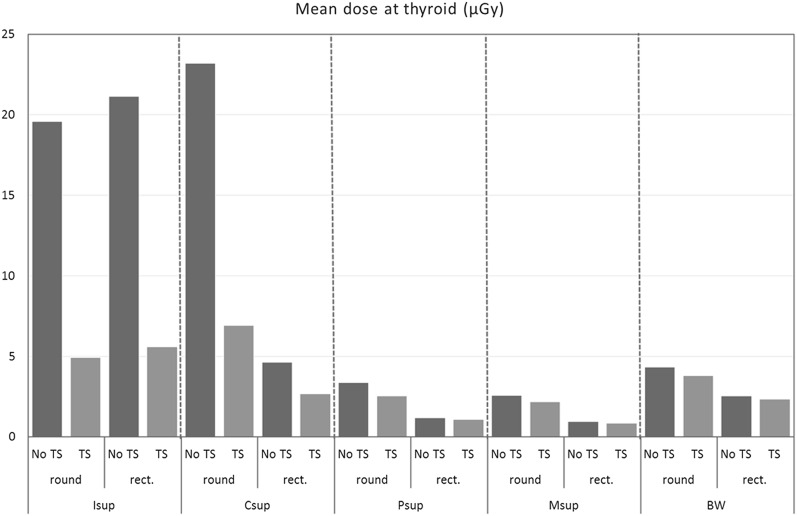
Results: The ANOVA test revealed that the differences between mean doses of all protocols and geometries were statistically significant, p < 0.001. For the Isup, thyroid dose levels were comparable with both collimators at a level indicating primary beam exposure. Thyroid shield reduced this dose with circa 75%. For the Csup position, round collimation also revealed primary beam exposure, and thyroid shield yield was 70%. In Csup with rectangular collimation, the thyroid dose was reduced with a factor 4 compared with round collimation and thyroid shield yielded an additional 42% dose reduction. The thyroid dose levels for the Csup, Psup, Msup and BW exposures were lower with rectangular collimation without thyroid shield than with round collimation with thyroid shield. With rectangular collimation, the thyroid shield in Psup, Msup and BW reduced the dose 10% or less, where dose levels were already low, implying no clinical significance.
Conclusions: For the exposures in the upper anterior region, thyroid shield results in an important dose reduction for the thyroid. For the other exposures, thyroid shield augments little to the reduction achieved by rectangular collimation. The use of thyroid shield is to be advised, when performing upper anterior radiography.
Keywords: dental radiography; radiation dosimetry; radiation protection; thyroid gland.
Figures
Similar articles
-
Kerma area product (KAP) and scatter measurements for intraoral X-ray machines using three different types of round collimation compared with rectangular beam limiter.Dentomaxillofac Radiol. 2019 Feb;48(2):20180183. doi: 10.1259/dmfr.20180183. Epub 2018 Nov 7. Dentomaxillofac Radiol. 2019. PMID: 30346798 Free PMC article.
-
Reducing the risk of intraoral radiographic imaging with collimation and thyroid shielding.Gen Dent. 2014 Jul-Aug;62(4):34-40. Gen Dent. 2014. PMID: 24983168
-
Effect of leaded glasses and thyroid shielding on cone beam CT radiation dose in an adult female phantom.Dentomaxillofac Radiol. 2013;42(6):20120260. doi: 10.1259/dmfr.20120260. Epub 2013 Feb 14. Dentomaxillofac Radiol. 2013. PMID: 23412460 Free PMC article.
-
Evidence on radiation dose reduction using rectangular collimation: a systematic review.Int Dent J. 2019 Apr;69(2):84-97. doi: 10.1111/idj.12411. Epub 2018 Jun 29. Int Dent J. 2019. PMID: 29959778 Free PMC article.
-
Outcomes of different radioprotective precautions in children undergoing dental radiography: a systematic review.Eur Arch Paediatr Dent. 2020 Aug;21(4):463-508. doi: 10.1007/s40368-020-00544-8. Epub 2020 Jun 16. Eur Arch Paediatr Dent. 2020. PMID: 32557182
Cited by
-
Knowledge, Attitude and Practice about radiation safety among the undergraduates in Eastern province dental college.J Pharm Bioallied Sci. 2021 Nov;13(Suppl 2):S1442-S1447. doi: 10.4103/jpbs.jpbs_248_21. Epub 2021 Nov 10. J Pharm Bioallied Sci. 2021. PMID: 35018006 Free PMC article.
-
The use of a thyroid shield for intraoral anterior oblique occlusal views-a risk-based approach.Dentomaxillofac Radiol. 2018 Jan;47(1):20170140. doi: 10.1259/dmfr.20170140. Epub 2017 Oct 27. Dentomaxillofac Radiol. 2018. PMID: 28891710 Free PMC article.
-
Operator safety during the acquisition of intraoral images with a handheld and portable X-ray device.Dentomaxillofac Radiol. 2018 Feb;47(3):20160410. doi: 10.1259/dmfr.20160410. Epub 2018 Feb 7. Dentomaxillofac Radiol. 2018. PMID: 29319336 Free PMC article.
-
Kerma area product (KAP) and scatter measurements for intraoral X-ray machines using three different types of round collimation compared with rectangular beam limiter.Dentomaxillofac Radiol. 2019 Feb;48(2):20180183. doi: 10.1259/dmfr.20180183. Epub 2018 Nov 7. Dentomaxillofac Radiol. 2019. PMID: 30346798 Free PMC article.
-
Korean dentists' perceptions and attitudes regarding radiation safety and protection.Dentomaxillofac Radiol. 2018 Feb;47(3):20170228. doi: 10.1259/dmfr.20170228. Epub 2018 Jan 31. Dentomaxillofac Radiol. 2018. PMID: 29236521 Free PMC article.
References
-
- International Commission on Radiation Protection. 1977 Recommendations of the International Commission on Radiation Protection, ICRP publication 26. Ann ICRP 1977; 1(3).
-
- ICRP. The 2007 Recommendations of the International Commission on Radiological Protection. ICRP publication 103. Ann ICRP 2007; 37(2–4). - PubMed
-
- Memon A, Godward S, Williams D, Siddique I, Al-Saleh K. Dental X-rays and the risk of thyroid cancer: a case-control study. Acta Oncol 2010; 49: 447–53. doi: http://dx.doi.org/10.3109/02841861003705778 - DOI - PubMed
-
- Rush ER, Thompson NA. Dental radiography technique and equipment: how they influence the radiation dose received at the level of the thyroid gland. Radiography 2007; 13: 214–20. doi: http://dx.doi.org/10.1016/j.radi.2006.03.002 - DOI
-
- Ludlow JB, Davies-Ludlow LE, White SC. Patient risk related to common dental radiographic examinations: the impact of 2007 International Commission on Radiological Protection recommendations regarding dose calculation. J Am Dent Assoc 2008; 139: 1237–43. doi: http://dx.doi.org/10.14219/jada.archive.2008.0339 - DOI - PubMed
Publication types
MeSH terms
LinkOut - more resources
Full Text Sources
Other Literature Sources
