Review
. 2016 Mar 24:11:29.
doi: 10.1186/s13023-016-0412-4.

Vogt-Koyanagi-Harada disease: review of a rare autoimmune disease targeting antigens of melanocytes

Affiliations
Review

Vogt-Koyanagi-Harada disease: review of a rare autoimmune disease targeting antigens of melanocytes

Marcelo Mendes Lavezzo et al. Orphanet J Rare Dis. .

Abstract

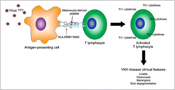
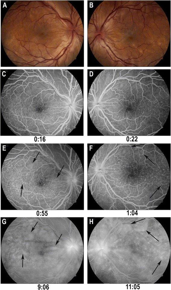
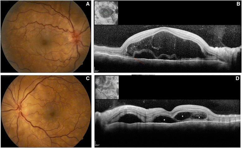
Vogt-Koyanagi-Harada disease (VKHD) is a rare granulomatous inflammatory disease that affects pigmented structures, such as eye, inner ear, meninges, skin and hair. This disease is mainly a Th1 lymphocyte mediated aggression to melanocytes after a viral trigger in the presence of HLA-DRB1*0405 allele. The absence of ocular trauma or previous intraocular surgery sets VKHD appart from sympathetic ophthalmia, its main differential diagnosis. The disease has an acute onset of bilateral blurred vision with hyperemia preceded by flu-like symptoms. The acute uveitic stage is characterized by a diffuse choroiditis with serous retinal detachment and optic disc hyperemia and edema. Fluorescein angiography in this phase demonstrates multiple early hyperfluorescent points. After the acute uveitic stage, ocular and integumentary system pigmentary changes may appear. Ocular findings may be accompanied by lymphocytic meningitis, hearing impairment and/or tinnitus in a variable proportion of patients. Prompt diagnosis followed by early, aggressive and long-term treatment with high-dose corticosteroids is most often ensued by good visual outcomes. However, some patients may experience chronic uveal inflammation with functional eye deterioration. The current review discusses the general features of VKHD, including epidemiology, classification into categories, differential diagnosis and current therapeutic approaches.

Keywords: Review; Uveitis; Vogt-Koyanagi-Harada disease.


Figures

Fig. 1
Fig. 1
Hypothetical scheme of VKHD pathogenesis. Courtesy of Arquivos Brasileiros de Oftalmologia – Damico, F.M., et al., New insights into Vogt-Koyanagi-Harada disease. Arq Bras Oftalmol, 2009. 72 [3]: p. 413-20 [39]
Fig. 2
Fig. 2
Acute uveitic stage: a and b: Right and left eye of a patient in the acute stage of Vogt-Koyanagi-Harada disease, presenting white- yellowish deep round lesions, hyperemia and blurring of the optic disc and exudative retinal detachment; c and d: Early fluorescein angiography, showing pinpoints and optic disc hyperfluorescence; e and f: Increase in pinpoints hyperfluorescence (arrows) and optic disc leakage; g and h: Coalescence of pinpoints hyperfluorescence resulting in pooling of the contrast (arrows) in exudative retinal detachment areas
Fig. 3
Fig. 3
OCT scans in the acute uveitic stage. a and c Fundoscopic aspect with optic disc swelling and hyperemia, besides multiple yellowish deep round lesions and exudative retinal detachment. b OCT scan showing a bullous unique exudative retinal detachment with hyperreflective material within the subretinal fluid (fibrin) and a strand continuous to the ellipsoid zone (arrow); d OCT scan of an exudative retinal detachment, with multiple compartments (asterisk) separated by membranous strutures
Fig. 4
Fig. 4
Right eye of a patient in the chronic stage. a: Fundoscopy with a mild depigmentation; b: OCT scan showing an increased choroidal thickness of 444 μm; c and d: Indocyanine green angiography showing multiple dark dots (arrows) and an uneven background choroidal fluorescence visible at the mid phase of ICGA
Fig. 5
Fig. 5
Poliosis of the lashes eyebrows and scalp hair
Fig. 6
Fig. 6
Vogt-Koyanagi-Harada disease treatment flowchart under consideration in the Uveitis Service, Hospital das Clínicas, Faculdade de Medicina, São Paulo University, São Paulo, SP, Brazil

References

    1. Moorthy RS, Inomata H, Rao NA. Vogt-Koyanagi-Harada syndrome. Surv Ophthalmol. 1995;39(4):265–292. doi: 10.1016/S0039-6257(05)80105-5. - DOI - PubMed
    1. Herbort CP, Mochizuki M. Vogt-Koyanagi-Harada disease: inquiry into the genesis of a disease name in the historical context of Switzerland and Japan. Int Ophthalmol. 2007;27(2–3):67–79. doi: 10.1007/s10792-007-9083-4. - DOI - PubMed
    1. Marmor MF, Ravin JG. The Artist’s Eyes - Vision and the History of Art. New York: Abrams; 2009. Chapter 34 - Goya’s Strange Malady; pp. 184–187.
    1. Rao NA, Gupta A, Dustin L, Chee SP, Okada AA, Khairallah M, et al. Frequency of distinguishing clinical features in Vogt-Koyanagi-Harada disease. Ophthalmology. 2010;117(3):591–599. doi: 10.1016/j.ophtha.2009.08.030. - DOI - PMC - PubMed
    1. Ohguro N, Sonoda KH, Takeuchi M, Matsumura M, Mochizuki M. The 2009 prospective multi-center epidemiologic survey of uveitis in Japan. Jpn J Ophthalmol. 2012;56(5):432–435. doi: 10.1007/s10384-012-0158-z. - DOI - PubMed

Publication types

MeSH terms