Case Reports
. 2016 Jun 1;25(11):2283-2294.
doi: 10.1093/hmg/ddw096. Epub 2016 Mar 22.

Mutations in C8ORF37 cause Bardet Biedl syndrome (BBS21)

Affiliations
Case Reports

Mutations in C8ORF37 cause Bardet Biedl syndrome (BBS21)

Elise Heon et al. Hum Mol Genet. .

Abstract

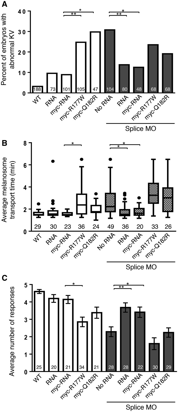
Bardet Biedl syndrome (BBS) is a multisystem genetically heterogeneous ciliopathy that most commonly leads to obesity, photoreceptor degeneration, digit anomalies, genito-urinary abnormalities, as well as cognitive impairment with autism, among other features. Sequencing of a DNA sample from a 17-year-old female affected with BBS did not identify any mutation in the known BBS genes. Whole-genome sequencing identified a novel loss-of-function disease-causing homozygous mutation (K102*) in C8ORF37, a gene coding for a cilia protein. The proband was overweight (body mass index 29.1) with a slowly progressive rod-cone dystrophy, a mild learning difficulty, high myopia, three limb post-axial polydactyly, horseshoe kidney, abnormally positioned uterus and elevated liver enzymes. Mutations in C8ORF37 were previously associated with severe autosomal recessive retinal dystrophies (retinitis pigmentosa RP64 and cone-rod dystrophy CORD16) but not BBS. To elucidate the functional role of C8ORF37 in a vertebrate system, we performed gene knockdown in Danio rerio and assessed the cardinal features of BBS and visual function. Knockdown of c8orf37 resulted in impaired visual behavior and BBS-related phenotypes, specifically, defects in the formation of Kupffer's vesicle and delays in retrograde transport. Specificity of these phenotypes to BBS knockdown was shown with rescue experiments. Over-expression of human missense mutations in zebrafish also resulted in impaired visual behavior and BBS-related phenotypes. This is the first functional validation and association of C8ORF37 mutations with the BBS phenotype, which identifies BBS21. The zebrafish studies hereby show that C8ORF37 variants underlie clinically diagnosed BBS-related phenotypes as well as isolated retinal degeneration.


Figures

Figure 1.
Figure 1.
Pedigree, C8ORF37 genomic structure and NMD assessment. (A) Schematic of genomic structure of two C8ORF37 isoforms with location of the novel mutation (K102*) hereby associated with BBS among the known mutations (associated with severe RP with maculopathy). (B) Pedigree showing first-degree consanguinity and family structure. Arrow: proband; −: mutant allele; +: WT allele; clear symbol: not affected; dark symbol: affected. (C) Semi-quantitative analysis of RT-PCR to assess the role of NMD. Products are cDNAs derived from lymphoblast cell lines from the patient with the variant c.304A> T and a WT individual for the c.304 position. The effect of treatments with cycloheximide (CHX) and dimethyl sulfoxide (DMSO) show that c.304>T undergoes NMD. Ladder indicates each 100 base pairs, with a strong band corresponding to 500 base pairs. PPIA served as a control.
Figure 2
Figure 2
Clinical phenotype at age 16 years. Upper panel: photograph of the retina of both eyes at the age of 16 years centered on the optic nerve (ON) and macula, the center of which is the fovea (f, dotted line). The paler area around the ON represents retinal atrophy and the foveal reflex is blunt. Middle panel: this is correlated by the OCT of left eye centered of the foveal. The foveal depression is reduced compared to the normal OCT, which corresponds to the blunt reflex on the photograph. The foveal depression is also thicker with the continuation of the GCL and IPL layers, which is diagnostic of a mild hypoplasia of the fovea. The resolution of the OCT allows the differentiation of the retinal layers, defined on the normal OCT; ILM/NFL: inner limiting membrane/nerve fiber layer; GCL: ganglion cell layer; IPL: inner plexiform layer; INL: inner nuclear layer; OPL: outer plexiform layer; INL: inner nuclear layer; OLM: outer limiting membrane; ISL: inner segment layer; CL: ciliary line; OSL: outer segment layer; RPE/BM: retinal pigment epithelium/Bruch’s membrane. The photoreceptor layer includes ISL, CL and OSL. D and lower panel: Goldmann visual fields (III4e isopter) show constriction OS (left eye) more than the right eye (OD). The dotted red line traces a normal field diameter of 120° (horizontal) × 100º (vertical).
Figure 3
Figure 3
Danio rerio as a model to study the functional roles of c8orf37. (A) Schematic presentation of genomic structure of c8orf37. The protein-coding region of c8orf37 has six exons. Translation start site is depicted with a curved arrow; the sites of ATG and splice MOs are shown with single and double bars below exons 1 and 2, respectively. The primers used for RT-PCR in (D) and (E) are flanked with black arrows. (B) Protein conservation between human (HS) and zebrafish (DR). The alignment was performed via ClustalO using the following protein sequences: Homo sapiens (HS) (NP_808880.1) and D. rerio (DR) (XP_003200284.2). There is evolutionary conservation of reported mutations in RP64 and CORD16 patients (p.Leu166, p.Arg177, p.Gln182 and p.Trp185) as well as our novel mutation site boxed in black: p.Lys102. Two missense mutation sites studied in this report (p.Arg177 and p.Gln182) are boxed in gray. The first amino acid of exon2 in HS and DR is highlighted in gray. (C) Partial alignment of C8ORF37 orthologs. The alignment that was performed with ClustalO shows evolutionary conservation of the novel mutation, p.Lys102, among human, rat (NP_001007747.1), mouse (NP_080281.3), chicken (XP_418346.2), zebrafish and frog (XP_002931575.1). (D) Temporal expression profiles of c8orf37 by RT-PCR. Robust expression of c8orf37 between 10 and 13 somite stages and 5 dpf enabled us to elucidate BBS-related and visual function phenotypes of c8orf37. β-actin served as a control. (E) Expression of c8orf37 in adult fish tissues by RT-PCR. The ubiquitous expression of c8orf37 was robust in the tissues tested. β-actin served as a control.
Figure 4
Figure 4
Knockdown of c8orf37 reveals extraocular BBS-related defects. (A) The KV phenotype was assessed at 10–13 somite stages evaluating the size of the vesicle (arrow) relative to the notochord and was abnormal in the ATG morphant. (B) Immunohistochemisty of KVs. The confocal images of KVs confirmed abnormal vesicle formation in the ATG morphants and suggested reduced number of motile cilia in it. Acetylated tubulin served as a marker for cilia and toPro-3 stained nuclei. (C) Graphic presentation of KV defects analyses. Abnormal KV formation, either reduced or absent, was observed in the both ATG and splice morphants in a MO-dose dependent manner. Data present number and percentage of embryos with KV defects. **P < 0.01 and ****P < 0.0001 with Fisher’s exact probability test. Number of larvae tested is in histogram bar. (D) Melanosome transport assay at 5 dpf in a control animal. After epinephrine shows the perinuclear distribution. To address the rate of retrograde intracellular trafficking, the time for a dark-adapted larva in epinephrine containing solution to retrieve melanosomes in the head area was measured. The time differences are showed in (E). (E) Graphic presentation of melanosome transport assay. Delayed melanosome transport was observed in both ATG and splice morphants in a MO-dose-dependent manner. *P < 0.05 with one-way ANOVA with Tukey adjustment. The splice morphants injected with 4 ng of MO also delayed trafficking significantly (P < 0.01) when compared to WT with Student t-test. The numbers of larvae tested are noted below lower whisker.
Figure 5
Figure 5
The vision startle responses. Graphic displays of the vision startle assay responses of the ATG and splice MO, both showing impaired visual function. Eight nanograms of Splice MO injection produced the most profound vision defects. Ctrl MO (normal) and crx MO (blind) were used as controls. Data present mean ± SEM. **P < 0.01 with one-way ANOVA with Tukey multiple comparison procedure. The number of animals studied is indicated in italics in the histogram bar.
Figure 6
Figure 6
Evaluation of missense mutations. (A) Graphic presentation of KV defects analyses. Both nascent form of synthetic RNA and myc-RNA produced normal KV formation in the splice morphants. Both the R177W and the Q182R mutant showed a KV defect. Mutant RNAs did not rescue the KV defects in the morphants but overexpression of either form induced significant KV malformation. Data present number and percentage of embryos with an abnormal KV. *P < 0.05 and **P < 0.01 with Fisher’s exact probability test. (B) Graphic presentation of melanosome transport assay. Delayed melanosome transport was normalized in the RNA or myc-RNA co-injected morphants. The data show that the R177W mutation itself induces a retrograde transport defect by delaying melanosome transport time. *P < 0.05 with one-way ANOVA with Tukey adjustment. The number of larvae tested is shown in each histogram bar. (C) Graphic presentation of the vision startle assays. Both RNA and myc-RNA were able to rescue the vision impairment in the splice morphants, while overexpression of the mutant form of RNAs failed to reverse the knockdown-driven vision impairments. In fact, the expression of the mutant RNAs alone resulted in significant vision defects. Data present mean ± SEM. *P < 0.05 and **P < 0.01 with one-way ANOVA with Tukey adjustment. The numbers of embryos studied are shown in the histogram bar.

References

    1. Beales P.L., Warner A.M., Hitman G.A., Thakker R., Flinter F.A. (1997) Bardet-Biedl syndrome: a molecular and phenotypic study of 18 families. J. Med. Genet., 34, 92–98. - PMC - PubMed
    1. Kerr E.N., Bhan A, Héon E. (2016) Exploration of the cognitive, adaptive and behavioral functioning of patients affected with Bardet-Biedl syndrome. Clin. Genet., 89, 426–433. - PubMed
    1. Kwitek-Black A.E., Carmi R., Duyk G.M., Buetow K.H., Elbedour K., Parvari R., Yandava C.N., Stone E.M., Sheffield V.C. (1993) Linkage of Bardet-Biedl syndrome to chromosome 16q and evidence for non-allelic genetic heterogeneity. Nat. Genet., 3, 392–396. - PubMed
    1. Leppert M., Baird L., Anderson K.L., Otterud B., Lupski J.R., Lewis R.A. (1994) Bardet-Biedl syndrome is linked to DNA markers on chromosome 11q and is genetically heterogeneous. Nat. Genet., 7, 108–112. - PubMed
    1. Sheffield V.C., Carmi R., Kwitek-Black A., Rokhlina T., Nishimura D., Duyk G.M., Elbedour K., Sunden S.L., Stone E.M. (1994) Identification of a Bardet-Biedl syndrome locus on chromosome 3 and evaluation of an efficient approach to homozygosity mapping. Hum. Mol. Genet., 3, 1331–1335. - PubMed

Publication types