. 2016 Jun;139(Pt 6):1649-56.
doi: 10.1093/brain/aww055. Epub 2016 Mar 23.

De novo PMP2 mutations in families with type 1 Charcot-Marie-Tooth disease

Affiliations

De novo PMP2 mutations in families with type 1 Charcot-Marie-Tooth disease

William W Motley et al. Brain. 2016 Jun.

Abstract

We performed whole exome sequencing on a patient with Charcot-Marie-Tooth disease type 1 and identified a de novo mutation in PMP2, the gene that encodes the myelin P2 protein. This mutation (p.Ile52Thr) was passed from the proband to his one affected son, and segregates with clinical and electrophysiological evidence of demyelinating neuropathy. We then screened a cohort of 136 European probands with uncharacterized genetic cause of Charcot-Marie-Tooth disease and identified another family with Charcot-Marie-Tooth disease type 1 that has a mutation affecting an adjacent amino acid (p.Thr51Pro), which segregates with disease. Our genetic and clinical findings in these kindred demonstrate that dominant PMP2 mutations cause Charcot-Marie-Tooth disease type 1.

Keywords: CMT; Charcot-Marie-Tooth disease; PMP2; myelin P2 protein; peripheral neuropathy.


Figures

None
Charcot-Marie-Tooth disease type 1 (CMT1) is a dominantly inherited demyelinating peripheral neuropathy with five known genetic causes. Motley et al. add to these by describing dominant de novo mutations in the gene encoding myelin P2 protein, PMP2, which co-segregate with CMT1 in two families.
Figure 1
Figure 1
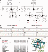
De novo PMP2 mutations segregate with disease in two families with CMT1. In Family 1, a c.155T > C mutation segregates with CMT1 and affects a conserved amino acid in the beta barrel of P2. (A) Whole exome sequencing of our proband Subject II.3 identified a c.155T > C substitution. A candidate screen for mutations in families with CMT identified a c.151A > C mutation in Family 2. The genotypes of individuals whose DNA was collected and Sanger sequenced is shown below pedigree symbols. In Family 1, neither parent has any clinical evidence of neuropathy and neither harbours the mutation. One of the proband’s sons is affected (Subject III.2) and harbours the mutation. In Family 2 the c.151A > C mutation segregates with CMT1 and affects a conserved amino acid adjacent to the mutation in Family 1. In Family 2, the proband (Subject III.3), her mother (Subject II.3), her two sisters (Subjects III.1 and III.2), and two nieces (Subjects IV.1 and IV.5), all show clinical features of neuropathy and carry the mutation, while the proband’s two aunts and one uncle do not have neuropathy and do not carry the mutation. The proband’s maternal grandparents are deceased, but did not show any signs of neuropathy. (B) Normal Sanger sequencing traces of the PMP2 gene are shown from the Family 1 proband’s parents, who are both unaffected, suggesting that the mutation arose de novo in our proband, and was passed onto his affected son. Sanger sequencing traces from the proband and her mother show the mutation in Family 2, while the proband’s unaffected uncle and aunt do not have the mutation. (C) ClustalW2 alignments of the myelin P2 protein from divergent species. The p.Ile43Asn, p.Thr51Pro, and p.Ile52Thr CMT1-associated variants disrupt amino acid residues that are conserved in mammals, but are not present in teleosts or invertebrates. (D) The crystal structure of the P2 protein is shown modelled with a bound palmitate. Helical structures are shown in light green and beta strands are shown in teal. The three CMT1-associated variants are shown, and they cluster in adjacent positions of beta strand B (p.Ile43Asn) and beta strand C (p.Thr51Pro, and the p.Ile52Thr).
Figure 2
Figure 2
Sural nerve biopsy from the proband in Family 1. Digital electron micrograph from the sural nerve biopsy of the proband (Patient II.3) at age 17. The density of myelinated axons is reduced, but actively degenerating myelinated axons were not seen. Some of the myelinated axons had myelin sheaths that were inappropriately thin for the axon diameter, and some rudimentary onion bulbs were seen; one of which is indicated in the boxed region. There is increased endoneurial collagen. The inset is a higher magnification image of the boxed region, showing a thinly myelinated axon surrounded by Schwann cell processes. Scale bar = 1 µm.

References

    1. Barrette B, Calvo E, Vallières N, Lacroix S. Transcriptional profiling of the injured sciatic nerve of mice carrying the Wld(S) mutant gene: identification of genes involved in neuroprotection, neuroinflammation, and nerve regeneration. Brain Behav Immun 2010; 24: 1254–67. - PubMed
    1. Berman HM, Westbrook J, Feng Z, Gilliland G, Bhat TN, Weissig H, et al. The protein data bank. Nucleic Acids Res 2000; 28: 235–42. - PMC - PubMed
    1. Chrast R, Saher G, Nave K-A, Verheijen MHG. Lipid metabolism in myelinating glial cells: lessons from human inherited disorders and mouse models. J Lipid Res 2011; 52: 419–34. - PMC - PubMed
    1. Collins JS, Schwartz CE. Detecting polymorphisms and mutations in candidate genes. Am J Hum Genet 2002; 71: 1251–2. - PMC - PubMed
    1. DeArmond SJ, Deibler GE, Bacon M, Kies MW, Eng LF. A neurochemical and immunocytochemical study of P2 protein in human and bovine nervous systems. J Histochem Cytochem 1980; 28: 1275–85. - PubMed

Publication types