. 2016 Jun 1;115(6):2937-49.
doi: 10.1152/jn.00671.2015. Epub 2016 Mar 23.

Biophysical constraints on lateral inhibition in the olfactory bulb

Affiliations

Biophysical constraints on lateral inhibition in the olfactory bulb

Alexa B R McIntyre et al. J Neurophysiol. .

Abstract

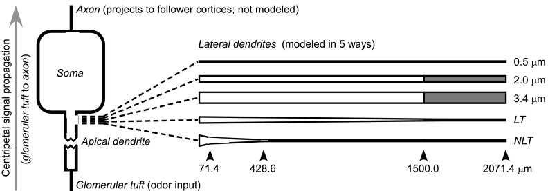
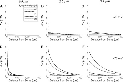
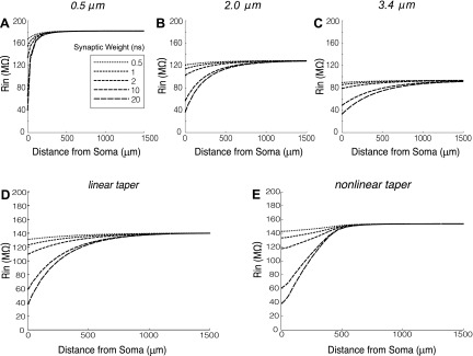
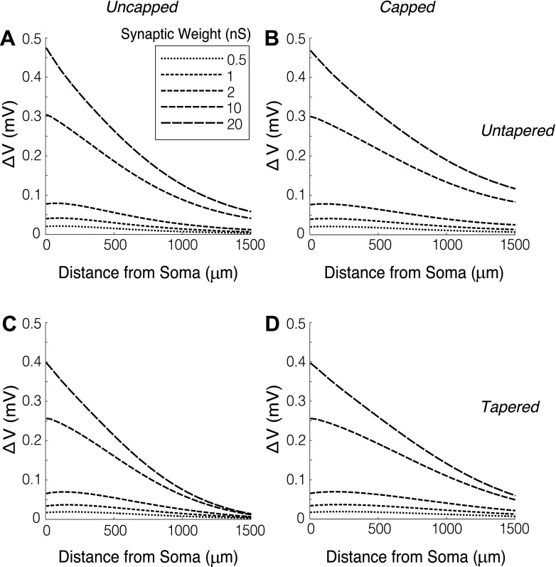
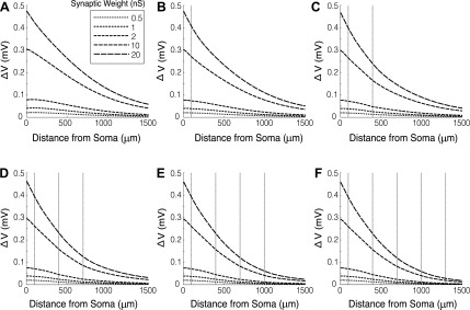
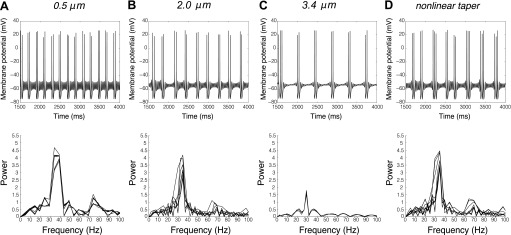
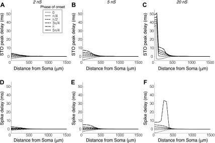
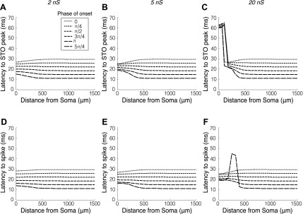
The mitral cells (MCs) of the mammalian olfactory bulb (OB) constitute one of two populations of principal neurons (along with middle/deep tufted cells) that integrate afferent olfactory information with top-down inputs and intrinsic learning and deliver output to downstream olfactory areas. MC activity is regulated in part by inhibition from granule cells, which form reciprocal synapses with MCs along the extents of their lateral dendrites. However, with MC lateral dendrites reaching over 1.5 mm in length in rats, the roles of distal inhibitory synapses pose a quandary. Here, we systematically vary the properties of a MC model to assess the capacity of inhibitory synaptic inputs on lateral dendrites to influence afferent information flow through MCs. Simulations using passivized models with varying dendritic morphologies and synaptic properties demonstrated that, even with unrealistically favorable parameters, passive propagation fails to convey effective inhibitory signals to the soma from distal sources. Additional simulations using an active model exhibiting action potentials, subthreshold oscillations, and a dendritic morphology closely matched to experimental values further confirmed that distal synaptic inputs along the lateral dendrite could not exert physiologically relevant effects on MC spike timing at the soma. Larger synaptic conductances representative of multiple simultaneous inputs were not sufficient to compensate for the decline in signal with distance. Reciprocal synapses on distal MC lateral dendrites may instead serve to maintain a common fast oscillatory clock across the OB by delaying spike propagation within the lateral dendrites themselves.

Keywords: cable theory; computational neuroscience; lateral dendrites; mitral cell; olfaction.


Figures

Fig. 1.
Fig. 1.
Model schematic. Afferent signal propagation in MCs begins in the glomerular tuft, proceeding through the apical dendrite and soma (in one of which MC spikes are initiated); these MC spikes then propagate down the axon (centripetally, to higher cortices) and the lateral dendrites (to effect lateral inhibition within the OB). Inhibitory synaptic inputs on these lateral dendrites thus fall outside of the axis of centripetal signal propagation, raising the question of whether and to what extent these synaptic inputs are able to modify olfactory signal propagation to the piriform cortex and other postbulbar structures. Five model dendrites were used to study this question: three of fixed diameter (0.5, 2.0, and 3.4 μm), one linearly tapering (LT), and one realistic, nonlinearly tapering (NLT; see materials and methods). Except where noted in the text, simulations were performed out to 2,071.4 μm (shaded region), but only analyzed out to 1,500 μm (nonshaded region) to avoid reflection effects due to dendritic caps (ends). The taper of the nonlinearly tapering dendrite changed at 71.4 and 428.6 μm (see materials and methods).
Fig. 2.
Fig. 2.
Changes in the membrane potential of the MC soma (ΔV) in response to inhibitory synaptic inputs along the lateral dendrite. Positive values on the ordinate denote hyperpolarization at the soma. The location of synaptic input ranged from 0 to 1,500 μm from the soma (abscissa). The maximum synaptic conductance was modeled as 0.5, 1, 2, 10, and 20 nS (inset); single synaptic events are estimated at 0.5–2.0 nS, with 10- to 20-nS conductances representing reasonable net inhibitory synaptic conductances that can be evoked in MCs by recurrent OB network activity (Schoppa 2006; Schoppa et al. 1998). Simulations were performed using two different chloride reversal potentials (A–C: −70 mV; D–F: −78 mV; see results for interpretations) and three lateral dendritic diameters (A and D: 0.5 μm; B and E: 2.0 μm; C and F: 3.4 μm). A diameter of 2.0 μm reflects the most proximal ∼40–80 μm of the lateral dendrite, whereas 0.5 μm better reflects its diameter more distally. 3.4 μm reflects the diameter of the primary dendrite and the immediate junction of the lateral dendrite.
Fig. 3.
Fig. 3.
Changes in somatic input resistance Rin with the opening of a shunting conductance along the mitral cell dendrite (abscissa). Five maximum synaptic conductances were modeled (inset). Rin was measured based on the change in membrane potential in response to a negative square pulse (ΔVpulse < 5 mV) applied to the soma under current clamp conditions. The reversal potential of the shunt was −70 mV, although adjustments to this parameter did not affect results. A: 0.5-μm diameter dendrite. B: 2.0-μm dendrite. C: 3.4-μm dendrite. D: linearly tapering dendrite with diameter tapering from 2 μm at the soma to 0.5 μm at 1,500-μm distance (then extending to 2,071.4 μm at a constant 0.5-μm diameter to avoid capping effects). E: nonlinearly tapering dendrite with diameter tapering from 3.4 μm at the soma to 2 μm at 71.4-μm distance, then to 0.5 μm at 428.6-μm distance, and then extending to 2,071.4 μm at a constant 0.5-μm diameter to avoid capping effects. See text for details.
Fig. 4.
Fig. 4.
Comparison of the change in membrane potential at the MC soma (ΔV) in linearly tapered and untapered dendrites, with or without capped ends, in response to inhibitory synaptic input. Synaptic reversal potential was −70 mV. A and B: 2-μm untapered dendrites. C and D: dendrites linearly tapered from 2 μm at the soma to 0.5 μm at 1,500 μm along the dendrite. A and C: uncapped dendrites, generated by simulating out to 2,071.4-μm distance (constant 0.5-μm diameter beyond 1,500-μm distance). B and D: dendrites capped at 1,500 μm, such that current reflects off of the capped end and marginally enhances efficacy at the soma. Note the change in scale on the ordinate compared with Fig. 1.
Fig. 5.
Fig. 5.
Comparison of the change in membrane potential at the MC soma (ΔV) in branched and unbranched lateral dendrites. Dendritic diameter was uniform (2 μm) in the main lateral dendrite and across all branches. The number of branches was increased from 0 to 5 (A to F, respectively), where dashed vertical lines indicate branch points. Branches did not provide additional synaptic inputs. Synaptic reversal potential was −70 mV.
Fig. 6.
Fig. 6.
Active cell model properties. A–D, top: somatic membrane potential time series in the active MC model alternate bursts of action potentials and interburst intervals exhibiting subthreshold oscillations. Bottom: power spectra of subthreshold oscillations within interburst intervals from each corresponding time series. MC lateral dendritic diameters were modeled at four diameters: 0.5 μm (A), 2.0 μm (B), 3.4 μm (C), and nonlinearly tapered from 3.4 to 0.5 μm (D), as described in Fig. 2E (see materials and methods). In all cases, activity was generated by 180 pA of depolarizing current injected into the soma.
Fig. 7.
Fig. 7.
Effects of inhibitory dendritic synaptic inputs on STO phase and spike timing in the active model MC. The lateral dendrite was nonlinearly tapered (Fig. 1, NLT; see also Figs. 2E and 5D). A–C: lead (negative) or lag (positive) in STO timing induced by inhibitory synaptic inputs delivered at six phases of the original MC STO (inset). A zero phase of onset indicates that the onset of the inhibitory postsynaptic current coincided with the somatic STO peak (phase of maximum depolarization). D–F: changes in spike timing induced by inhibitory synaptic inputs delivered at six phases of the MC STO immediately preceding the onset of the first spike of a burst. Inputs had a reversal potential of −70 mV and were modeled at three synaptic weights (peak synaptic conductances): 2 nS (A and D), 5 nS (B and E), or 20 nS (C and F). Phases of onset beyond 5π/4 were excluded from the plots as STO peaks were truncated, or spikes skipped (i.e., delayed for at least a full cycle), depending on the strength of input.
Fig. 8.
Fig. 8.
MC spike timing regulation by inhibitory synaptic inputs and intrinsic STO dynamics. Panels depict the same results shown in Fig. 6, but highlight the constraining effects on MC spike timing. A–C: latency between the onset of an inhibitory input delivered to a cell during a nonspiking STO period and the following STO peak. Very strong synaptic inputs delivered adjacent to the soma could induce rebound spikes, substantially delaying the following STO (C). D–F: latency between the onset of an inhibitory input delivered immediately preceding the onset of a spike burst and the first spike. Multiple phases of onset of the inhibitory synaptic input were tested (inset). Inputs had a reversal potential of −70 mV and were modeled at three synaptic weights (peak synaptic conductances): 2 nS (A and D), 5 nS (B and E), or 20 nS (C and F). Phases of onset were defined with 0 as the peak of the preceding STO and 2π as the peak of the following STO or spike in the absence of synaptic input. Phases of onset beyond 5π/4 were excluded from the plots as STO peaks were truncated, or spikes skipped (i.e., delayed for at least a full cycle), depending on the strength of input. Strong inhibitory inputs between two STOs were also capable of triggering rebound spikes and delaying the following STO peak (e.g., C). The convergence of the six curves as proximity to the soma increases reflects the degree to which MC STOs are reset to a common phase by inhibitory synaptic input (Rubin and Cleland, 2006).

Similar articles

Cited by

References

    1. Arruda-Carvalho M, Akers KG, Guskjolen A, Sakaguchi M, Josselyn SA, Frankland PW. Posttraining ablation of adult-generated olfactory granule cells degrades odor-reward memories. J Neurosci 34: 15793–15803, 2014. - PMC - PubMed
    1. Balu R, Larimer P, Strowbridge BW. Phasic stimuli evoke precisely timed spikes in intermittently discharging mitral cells. J Neurophysiol 92: 743–753, 2004. - PubMed
    1. Bartel D, Rela L, Hsieh L, Greer C. Dendrodendritic synapses in the mouse olfactory bulb external plexiform layer. J Comp Neurol 523: 1145–1161, 2015. - PMC - PubMed
    1. Bazhenov M, Rulkov N, Timofeev I. Effect of synaptic connectivity on long-range synchronization of fast cortical oscillations. J Neurophysiol 100: 1562–1575, 2008. - PMC - PubMed
    1. Ben-Ari Y. Excitatory actions of GABA during development: the nature of the nurture. Nat Rev Neurosci 3: 728–739, 2002. - PubMed

Publication types

Substances

LinkOut - more resources