. 2016 Sep;18(9):1242-52.
doi: 10.1093/neuonc/now043. Epub 2016 Mar 23.

Autocrine VEGFR1 and VEGFR2 signaling promotes survival in human glioblastoma models in vitro and in vivo

Affiliations

Autocrine VEGFR1 and VEGFR2 signaling promotes survival in human glioblastoma models in vitro and in vivo

Emese Szabo et al. Neuro Oncol. 2016 Sep.

Abstract

Background: Although the vascular endothelial growth factor (VEGF)/VEGF receptor (VEGFR) system has become a prime target for antiangiogenic treatment, its biological role in glioblastoma beyond angiogenesis has remained controversial.

Methods: Using neutralizing antibodies to VEGF or placental growth factor (PlGF) or the tyrosine kinase inhibitor, cediranib, or lentiviral gene silencing, we delineated autocrine signaling in glioma cell lines. The in vivo effects of VEGFR1 and VEGFR2 depletion were evaluated in orthotopic glioma xenograft models.

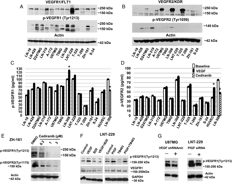
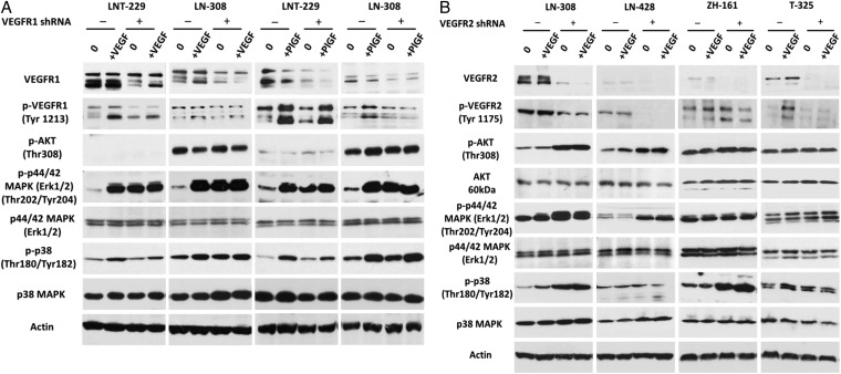
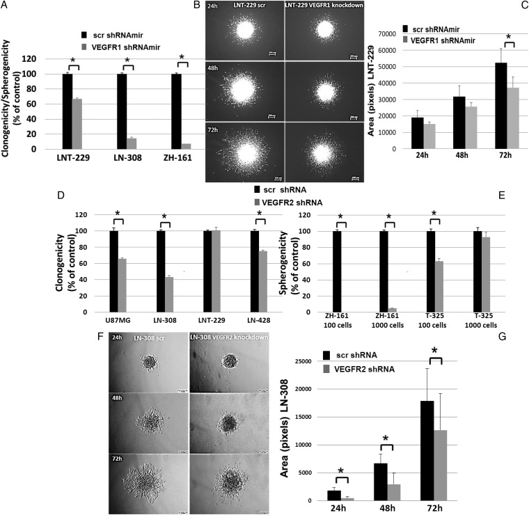
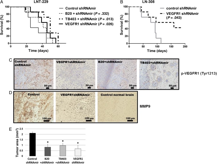
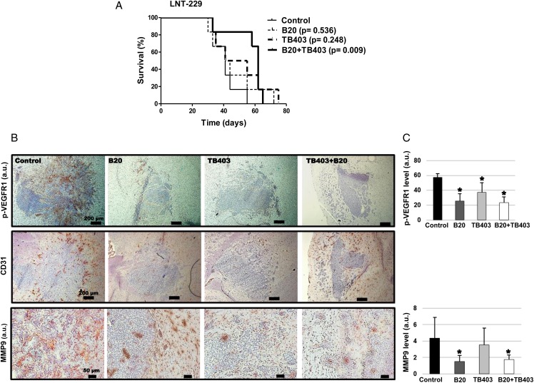
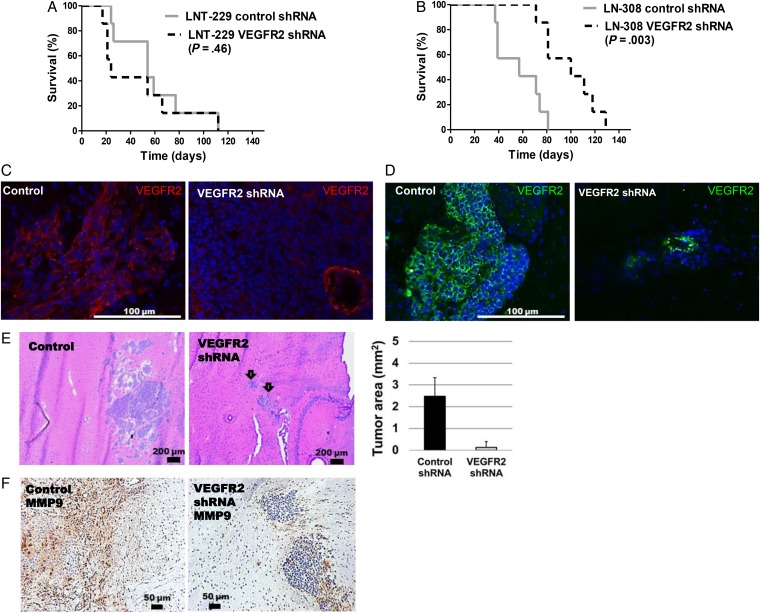
Results: VEGFR1 and VEGFR2 modulated glioma cell clonogenicity, viability, and invasiveness in vitro in an autocrine, cell-line-specific manner. VEGFR1 silencing promoted mitogen-activated protein kinase/extracellular signal-regulated kinase (MAPK/ERK) signaling, whereas VEGFR2 silencing resulted in cell-type dependent activation of the protein kinase B (PKB)/AKT and MAPK/ERK pathways. These responses may represent specific escape mechanisms from VEGFR inhibition. The survival of orthotopic glioma-bearing mice was prolonged upon VEGFR1 silencing in the LNT-229, LN-308, and U87MG models and upon VEGFR2 silencing in LN-308 and U87MG. Disruption of VEGFR1 and VEGFR2 signaling was associated with decreased tumor size, increased tumor necrosis, or loss of matrix metalloproteinase 9 (MMP9) immunoreactivity. Neutralizing VEGF and PlGF by specific antibodies was superior to either antibody treatment alone in the VEGFR1-dependent LNT-229 model.

Conclusions: Differential dependence on autocrine signaling through VEGFR1 and VEGFR2 suggests a need for biomarker-stratified VEGF(R)-based therapeutic approaches to glioblastoma.

Keywords: PlGF; VEGF; angiogenesis; glioblastoma; signaling.


Figures

Fig. 1.
Fig. 1.
Autocrine and induced vascular endothelial growth factor receptor (VEGFR) activation in glioma cells. (A, B) The levels of total VEGFR1 and VEGFR2 as well as of phosphorylated VEGFR1Tyr1213 or VEGFR2Tyr1059 were assessed by immunoblot, using actin as a loading control. (C, D) Constitutive and VEGF-evoked total phosphorylation levels of VEGFR1 or VEGFR2 were determined by ELISA (*P < .05). (E) VEGFR1 and VEGFR2 phosphorylation in response to increasing concentrations of cediranib (2 h) in ZH-161 cells were detected by immunoblot. (F) Effects of VEGF or PlGF stimulation or neutralizing anti-VEGF (B20) or anti-placental growth factor (PlGF) (TB403) antibodies on VEGFR1Tyr1213 in LNT-229 cells were assessed by immunoblot; cells were incubated for 15 minutes with VEGF (500 ng/mL) or PlGF(1 + 2) (200 ng/mL) alone or in combination with neutralizing VEGF or PlGF antibodies (100 µg/mL). (G) Effects of lentivirus-mediated VEGFA shRNAmir (left) or PlGF siRNA (right) on constitutive p-VEGFR1Tyr1213 in U87MG or LNT-229 cells, respectively, were evaluated by immunoblot.
Fig. 2.
Fig. 2.
Altered downstream signaling in vascular endothelial growth factor receptor (VEGFR)1- and VEGFR2-depleted glioma cells. (A) Stably VEGFR1 gene-silenced LNT-229 or LN-308 cells or (B) VEGFR2 gene-silenced LN-308, LN-428, ZH-161 or T-325 cells, or corresponding controls were assayed for changes in downstream signaling by immunoblot. After 12 hours of serum starvation, subconfluent cells were untreated or stimulated with VEGF (500 ng/mL) or placental growth factor (PlGF) (1 + 2) (100 + 100 ng/mL) as indicated for 15 minutes.
Fig. 3.
Fig. 3.
Biological effects of vascular endothelial growth factor receptor (VEGFR) signaling inhibition in glioma cells. Effects of VEGFR1 gene silencing on (A) clonogenicity or spherogenicity and (B, C) invasion of LNT-229 were studied. (D, E) Effects of VEGFR2 gene silencing on clonogenicity and spherogenicity was evaluated. (F, G). Invasiveness of VEGFR2-depleted LN-308 cells was assessed by spheroid invasion assays. The data represent the average fold change in area of 3 spheroids ± standard deviation (*P < .05).
Fig. 4.
Fig. 4.
Genetic depletion of vascular endothelial growth factor receptor (VEGFR)1 or pharmaceutical neutralization of both VEGFR1 ligands, VEGF and placental growth factor (PlGF), delay tumor growth in vivo. (A) 75 000 nontargeting control shRNAmir-expressing human LNT-229 glioma cells were implanted into the brains of nude mice. The mice were treated either twice weekly with 20 mg/kg/day Xolair IgG control or 5 mg/kg/day B20 or 20 mg/kg/day TB403 by intraperitoneal injection. The treatment was initiated at the day of tumor implantation and maintained until the onset of clinical grade 2 symptoms. A parallel group of mice was transplanted with 75 000 VEGFR1-targeted shRNA expressing LNT-229 glioma cells. (B) 100 000 VEGFR1-silenced or corresponding control LN-308 cells were implanted into the brains of nude mice. Seven animals per group were used to monitor survival (Mantel-Cox test). (C) LNT-229 tumor specimens obtained per randomization list from animals sacrificed on the same day in each group when the first animal(s) became symptomatic were stained for p-VEGFR1Tyr1213 (brown color). Sections were counterstained with hematoxylin (blue). A higher magnification image shown in Fig. S12A confirmed subcellular localization. (D) LNT-229 tumor specimens or normal brain were stained for MMP9 immunoreactivity (brown color). Sections were counterstained with hematoxylin (blue). (E) LNT-229 tumor sizes were assessed on H&E-stained sections (n = 3; *P < .05, t test).
Fig. 5.
Fig. 5.
Synergistic growth inhibition by targeting both vascular endothelial growth factor receptor (VEGFR)1 ligands, VEGF and placental growth factor (PlGF) in vivo. (A) A similar experiment as in Fig. 4A was performed, but with the modification that antibody treatment was delayed until day 15 after tumor implantation and that another group of animals treated with both VEGF and PlGF antibody were included. (B) Tumor specimens obtained per randomization list from animals sacrificed on the same day in each group when the first animal(s) became symptomatic were stained for p-VEGFR1Tyr1213 (upper row, brown color), CD31 (middle row) or MMP9 (lower row). Sections were counterstained with hematoxylin (blue). (C) Quantification of immunoreactivity (n = 3; *P < .05, t test).
Fig. 6.
Fig. 6.
Genetic depletion of vascular endothelial growth factor receptor (VEGFR)2 delays tumor growth in the LN-308 but not in the LN-229 glioma model. (A,B) 75 000 LNT-229 or 100 000 LN-308 depleted of VEGFR2 or their shRNA control cells were implanted intracranially and monitored for survival (n = 7). (C, D) VEGFR2 gene silencing was confirmed by immunofluoresence microscopy at days 21 (LNT-229) or 37 (LN-308). Preservation of blood vessel labeling after tumor-specific gene silencing serves as an internal control. (E) LN-308 tumor sizes were determined based on H&E-stained sections (n = 3). (F) IHC for MMP9 levels upon VEGFR2 silencing. The mean area (a.u.) of MMP9-positive segments was reduced by ∼86% relative to control values (n = 3, P < .05).

References

    1. Folkins C, Shked Y, Man S et al. . Glioma tumor stem-like cells promote tumor angiogenesis and vasculogenesis via vascular endothelial growth factor and stromal-derived factor 1. Cancer Res. 2009;69 (18):7243–7251. - PMC - PubMed
    1. Plate KH, Breier G, Weich HA et al. . Vascular endothelial growth-factor and glioma angiogenesis - coordinate induction of VEGF receptors, distribution of VEGF protein and possible in-vivo regulatory mechanisms. Int J Cancer. 1994;59 (4):520–529. - PubMed
    1. Yao J, Wu XM, Zhuang GL et al. . Expression of a functional VEGFR-1 in tumor cells is a major determinant of anti-PlGF antibodies efficacy. Proc Natl Acad Sci U S A. 2011;108 (28):11590–11595. - PMC - PubMed
    1. Nomura M, Yamagishi S, Harada S et al. . Placenta growth factor (PlGF) mRNA expression in brain tumors. J Neurooncol. 1998;40 (2):123–130. - PubMed
    1. Carmeliet P, Moons L, Luttun A et al. . Synergism between vascular endothelial growth factor and placental growth factor contributes to angiogenesis and plasma extravasation in pathological conditions. Nat Med. 2001;7 (5):575–583. - PubMed

Publication types

Substances