. 2016 Mar 23;8(331):331ra41.
doi: 10.1126/scitranslmed.aad4583.

Targeting protein homeostasis in sporadic inclusion body myositis

Affiliations

Targeting protein homeostasis in sporadic inclusion body myositis

Mhoriam Ahmed et al. Sci Transl Med. .

Abstract

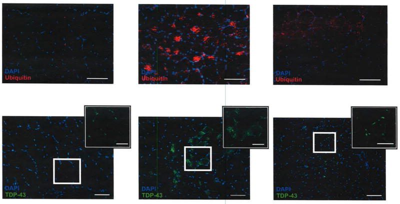
Sporadic inclusion body myositis (sIBM) is the commonest severe myopathy in patients more than 50 years of age. Previous therapeutic trials have targeted the inflammatory features of sIBM but all have failed. Because protein dyshomeostasis may also play a role in sIBM, we tested the effects of targeting this feature of the disease. Using rat myoblast cultures, we found that up-regulation of the heat shock response with arimoclomol reduced key pathological markers of sIBM in vitro. Furthermore, in mutant valosin-containing protein (VCP) mice, which develop an inclusion body myopathy, treatment with arimoclomol ameliorated disease pathology and improved muscle function. We therefore evaluated arimoclomol in an investigator-led, randomized, double-blind, placebo-controlled, proof-of-concept trial in sIBM patients and showed that arimoclomol was safe and well tolerated. Although arimoclomol improved some IBM-like pathology in the mutant VCP mouse, we did not see statistically significant evidence of efficacy in the proof-of-concept patient trial.


Conflict of interest statement

LG became an unpaid consultant to Orphazyme Aps (the owner of Arimoclomol) after completing this study. The other authors declare no competing interests.

Figures

Fig. 1
Fig. 1. β-APP overexpression or exposure to inflammatory mediators induces sIBM-like pathology in cultured rat myocytes that is abrogated by Arimoclomol
Shown is formation of cytoplasmic inclusion bodies (white arrows) in rat myocytes transfected with full-length human β-APP immunoreactive for (A) β-APP and ubiquitin and (B) TDP-43 and ubiquitin. (C) The number of myocytes containing ubiquitinated inclusion bodies as a percentage of the total number of myocytes present (n=3; p<0.05; one-way ANOVA). (D) Expression of TDP-43 (green) following empty vector (EV) or β-APP transfection and Arimoclomol treatment, and (E) the number of myocytes with cytoplasmic mislocalisation of TDP-43 (n=3, p<0.001; unpaired t-test). (F) TDP-43 expression (green) following exposure to inflammatory mediators and Arimoclomol, and (G) quantification of TDP-43 mislocalisation in cytokine-treated cultures (n=3, p<0.05; unpaired t-test). (H) Western blot analysis of TDP-43 expression in rat myocyte cultures exposed to cytokines in the presence and absence of Arimoclomol. Images show the expression of NF-κB subunit p65 (green) in (I) β-APP transfected cultures (DAPI labelled nuclei in blue), and (J) cultures exposed to cytokines in the presence and absence of Arimoclomol. (K) The number of rat myocytes with nuclear NF-κB subunit p65 as a percentage of the total number of myocytes present (n=3, p<0.05; one-way ANOVA). Error bars = S.E.M; scale bars: A, B =10 μm, D, I and J =20 μm. * indicates statistical significance.
Fig. 2
Fig. 2. Arimoclomol augments HSP70 expression and improves survival of cultured rat myocytes
(A) HSP70 expression (red) in cultured rat myocytes following empty vector (EV) transfection (left hand panel), β-APP overexpression (middle panel), or β-APP overexpression plus treatment with Arimoclomol (right hand panel). (B) HSP70 expression (red) in cultured rat myocytes that were untreated (control; left hand panel), or after exposure to IL-1β alone (middle panel) or IL-1β plus Arimoclomol (right hand panel). (C) Western blot analysis of HSP70 expression in rat myocyte cultures exposed to inflammatory mediators or after β-APP overexpression in the presence or absence of Arimoclomol. (D) Cytotoxicity in β-APP overexpressing rat myocyte cultures as a percentage of that in control cultures (n=3, p<0.02; one-way ANOVA), as assessed by a lactate dehydrogenase (LDH) assay. (E) Cytotoxicity following exposure to inflammatory mediators in the presence or absence of Arimoclomol (n=3, p<0.05; one-way ANOVA), as assessed by an MTT (3-(4, 5-dimethylthiazolyl-2)-2,5-diphenyltetrazolium bromide) assay. Error bars= S.E.M.; Scale bars=10 μm. * indicates statistical significance.
Fig. 3
Fig. 3. Disruption of protein homeostasis in rat myocyte cultures is prevented by Arimoclomol
(A) The cytosolic calcium ion response induced by the ER stressor thapsigargin (an indicator of ER stress) in β-APP overexpressing rat myocyte cultures and in cell cultures exposed to inflammatory mediators (n=3, p<0.05; one-way ANOVA). (B) Expression of the ER stress mediator CHOP determined by Western blot analysis in rat myocyte cultures overexpressing β-APP or exposed to inflammatory mediators, in the presence or absence of Arimoclomol. (C) The images show the presence of a cytoplasmic aggregate immunoreactive for p62 (red; white arrow) in a desmin-positive rat myocyte following β-APP transfection. (D) The images show the expression of the autophagic protein LC3-II (red; white arrow) in β-APP transfected desmin-positive rat myocytes after Arimoclomol treatment. The white arrow indicates punctate LC3-II staining of autophagosomes. (E) The chymotrypsin-like proteasome activity assessed 48 hours after β-APP transfection in the presence or absence of Arimoclomol (n=3, p<0.05; one-way ANOVA). (F) The expression of the autophagosome marker LC3 II in β-APP transfected rat myocytes with or without Arimoclomol (n=3, p<0.05; one-way ANOVA). EV, empty vector. Error bars=S.E.M; Scale bars=10 μm. * indicates statistical significance.
Fig. 4
Fig. 4. Arimoclomol treatment improves muscle strength, muscle contractile characteristics and IBM-like pathology in mutant VCP mice
(A) The change in grip strength in wildtype VCP (WT-VCP), mutant VCP (mVCP) and Arimoclomol-treated mutant VCP (mVCP+Ari) mice between 4 and 14 months (n=10; p<0.0001; unpaired t-test). (B) Typical traces of muscle twitch and maximum tetanic force of extensor digitorum longus (EDL) muscles in untreated and Arimoclomol-treated mutant VCP mice. (C) Mean maximum force of extensor digitorum longus muscles of WT-VCP, mutant VCP and Arimoclomol-treated mutant VCP mice (n=10; p<0.04; one-way ANOVA). (D) H&E staining of tibialis anterior (TA) muscles of mice in each experimental group (white arrow, atrophied fibre; black arrowheads, hypertrophic fibres). (E) H&E staining showing clear inflammatory cell infiltration in mutant VCP tibialis anterior muscle. Western blots show (F) MHC-1 and (G) phospho-IκBα expression in tibialis anterior muscles of mice in each experimental group. (H) Transmission electron microscopy of tibialis anterior muscles of mice in each experimental group. (I) Muscle fibre diameter in tibialis anterior muscles from untreated and Arimoclomol-treated mutant VCP mice compared to WT-VCP mice (n=3; p<0.0001; one-way ANOVA). (J) Western blot analysis of HSP70 expression in tibialis anterior muscles from mice in each experimental group. Bar chart shows mean relative optical density (n=3; p=0.01; unpaired t-test). Error bars = S.E.M, Scale bars: D, E =50 μm; H=2μm. * indicates statistical significance.
Fig. 5
Fig. 5. Arimoclomol abrogates IBM-like pathology in mutant VCP mouse muscle
Cross sections of tibialis anterior muscles from wildtype VCP (WT-VCP), mutant VCP (mVCP) and Arimoclomol-treated mutant VCP (mVCP+Ari) mice, immunostained for ubiquitin (red; A-C) and TDP-43 (green; D-F); nuclei stained with DAPI (blue). Scale bar = 50 μm; inserts = 25 μm.
Fig. 6
Fig. 6. Clinical trial secondary outcomes (efficacy measures)
The change from baseline to endpoint on 3 different clinical scales assessed at 4, 8 and 12 months (M) (mean ± S.E.M) in sIBM patients treated with Arimoclomol for 4 months. (A) IBMFRS score, (B) MMT average score, and (C) right hand grip MVICT score. The IBMFRS is a disease-specific functional questionnaire for patients with sIBM and measures physical function/disability. MMT is a measure of muscle strength scored by the physician based on the clinical assessment. MVICT is a measure of muscle strength performed using a quantitative muscle assessment system that uses an adjustable cuff to attach the patient’s arm or leg to an inelastic strap that is connected to a force transducer. Error bars = S.E.M. No statistically significant clinical efficacy measures were observed.

References

    1. Dimachkie MM, Barohn RJ. Inclusion body myositis. Current neurology and neuroscience reports. 2013;13(1):321. - PubMed
    1. Machado P, Brady S, Hanna MG. Update in inclusion body myositis. Current opinion in rheumatology. 2013;25(6):763–71. - PMC - PubMed
    1. Amato AA, Gronseth GS, Jackson CE, Wolfe GI, Katz JS, Bryan WW, Barohn RJ. Inclusion body myositis: clinical and pathological boundaries. Ann Neurol. 1996;40(4):581–6. - PubMed
    1. Amato AA, Barohn RJ. Evaluation and treatment of inflammatory myopathies. J Neurol Neurosurg Psychiatry. 2009;80(10):1060–8. - PubMed
    1. Cox FM, Titulaer MJ, Sont JK, Wintzen AR, Verschuuren JJ, Badrising UA. A 12-year follow-up in sporadic inclusion body myositis: an end stage with major disabilities. Brain. 2011;134(Pt 11):3167–75. - PubMed

Publication types

MeSH terms