. 2016 Aug 1;311(2):F330-42.
doi: 10.1152/ajprenal.00002.2016. Epub 2016 Mar 23.

Renal tubular SGK1 deficiency causes impaired K+ excretion via loss of regulation of NEDD4-2/WNK1 and ENaC

Affiliations

Renal tubular SGK1 deficiency causes impaired K+ excretion via loss of regulation of NEDD4-2/WNK1 and ENaC

Lama Al-Qusairi et al. Am J Physiol Renal Physiol. .

Abstract

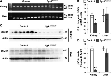
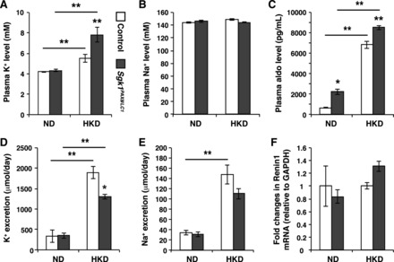
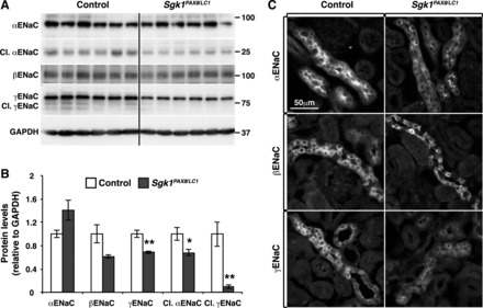
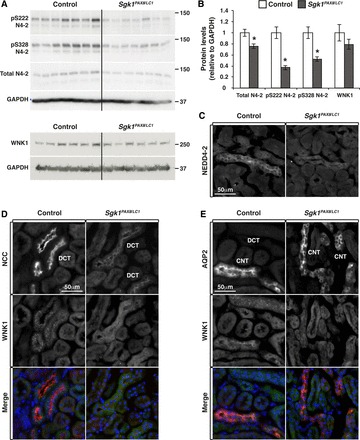
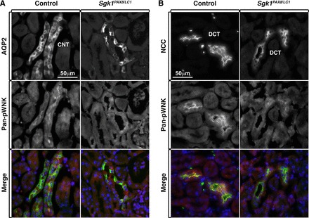
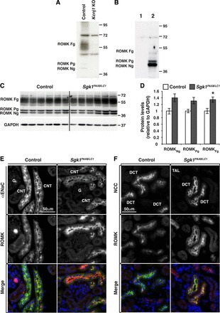
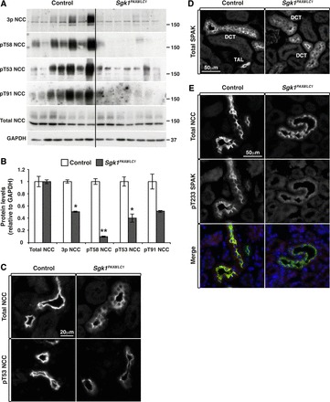
The stimulation of postprandial K(+) clearance involves aldosterone-independent and -dependent mechanisms. In this context, serum- and glucocorticoid-induced kinase (SGK)1, a ubiquitously expressed kinase, is one of the primary aldosterone-induced proteins in the aldosterone-sensitive distal nephron. Germline inactivation of SGK1 suggests that this kinase is fundamental for K(+) excretion under conditions of K(+) load, but the specific role of renal SGK1 remains elusive. To avoid compensatory mechanisms that may occur during nephrogenesis, we used inducible, nephron-specific Sgk1(Pax8/LC1) mice to assess the role of renal tubular SGK1 in K(+) regulation. Under a standard diet, these animals exhibited normal K(+) handling. When challenged by a high-K(+) diet, they developed severe hyperkalemia accompanied by a defect in K(+) excretion. Molecular analysis revealed reduced neural precursor cell expressed developmentally downregulated protein (NEDD)4-2 phosphorylation and total expression. γ-Epithelial Na(+) channel (ENaC) expression and α/γENaC proteolytic processing were also decreased in mutant mice. Moreover, with no lysine kinase (WNK)1, which displayed in control mice punctuate staining in the distal convoluted tubule and diffuse distribution in the connecting tubule/cortical colleting duct, was diffused in the distal convoluted tubule and less expressed in the connecting tubule/collecting duct of Sgk(Pax8/LC1) mice. Moreover, Ste20-related proline/alanine-rich kinase phosphorylation, and Na(+)-Cl(-) cotransporter phosphorylation/apical localization were reduced in mutant mice. Consistent with the altered WNK1 expression, increased renal outer medullary K(+) channel apical localization was observed. In conclusion, our data suggest that renal tubular SGK1 is important in the regulation of K(+) excretion via the control of NEDD4-2, WNK1, and ENaC.

Keywords: aldosterone; epithelial Na+ channel; epithelial transport; neural precursor cell expressed developmentally downregulated protein 4-2; phosphorylation; potassium; serum- and glucocorticoid-induced kinase 1; ubiquitylation; with no lysine kinase 1.


Figures

Fig. 1.
Fig. 1.
Serum- and glucocorticoid-induced kinase (Sgk)1 mRNA and SGK1 protein levels are decreased in the kidney but not in the liver of Sgk1Pax8/LC1 mutant mice. A: PCR on genomic DNA extracted from the total kidney and liver showing wild-type (WT) and mutant [knockout (KO)] Sgk1 bands after 12 days of doxycycline treatment. The intensity of the mutant band in the kidney was more pronounced than in the liver of the same animals. B: quantitative RT-PCR (TaqMan) analysis of Sgk1 mRNA in control and Sgk1Pax8/LC1 KO mice. A significant decreased of the Sgk1 mRNA level occurred in the kidney but not in the liver of the same animals (n = 7 control mice and 6 Sgk1Pax8/LC1 mice). C: Western blot (WB) analysis of phosphorylated (p)SGK1 (pThr256) in protein lysates from the total kidney (top) and liver (bottom) in control and Sgk1Pax8/LC1 KO mice. Actin was used as a loading control. D: protein quantification of the results in C showing an almost complete loss of pSGK1 in the kidney of mutant mice compared with control mice with no alteration of pSGK1 levels in the liver (n = 6 control mice and 7 Sgk1Pax8/LC1 mice). *P < 0.05; **P < 0.01.
Fig. 2.
Fig. 2.
K+ excretion is defective in Sgk1Pax8/LC1 mice under a high-K+ diet (HKD) but not a normal diet (ND). A and B: plasma K+ (A) and Na+ (B) in control (open bars) and Sgk1Pax8/LC1 (solid mice) mice showing significant hyperkalemia with a slight decrease in natremia in Sgk1Pax8/LC1 mice under the HKD (n = 5 control mice and 7 Sgk1Pax8/LC1 mice) but not under a ND (n = 12 control mice and 11 Sgk1Pax8/LC1 mice). C: plasma aldosterone was increased in Sgk1Pax8/LC1 versus control mice under a ND (n = 12 control mice and 13 Sgk1Pax8/LC1 mice) and under a HKD (n = 14 control mice and 11 Sgk1Pax8/LC1 mice). D and E: 24-h urinary K+ (D) and Na+ (E) excretion indicating reduced K+ excretion in Sgk1Pax8/LC1 mutant animals with no significant difference in Na+ excretion (n = 12 control mice and 13 Sgk1Pax8/LC1 mice). *α value < 0.05; **α value < 0.01. F: quantitative RT-PCR (Taqman) on RNA isolated from kidneys of control or Sgk1Pax8/LC1 mice kept under a ND or 48 h of a HKD.
Fig. 3.
Fig. 3.
Deregulation of epithelial Na+ channels (ENaC) in Sgk1Pax8/LC1 mice after 2 days of HKD. A: WB analysis of ENaC in control and Sgk1Pax8/LC1 mice showing a decrease in cleaved (Cl.) αENaC and in full-length and cleaved γENaC without an alteration in βENaC. B: protein quantification of the results in A (n = 6 control mice and 7 Sgk1Pax8/LC1 mice). C: immunofluorescence (IF) images of ENaC in control and Sgk1Pax8/LC1mutant mice showing a decrease in the apical localization of αENaC (top) and in the total expression of γENaC (bottom). βENaC exhibited a similar pattern in both genotypes (middle). *P < 0.05; **P < 0.01.
Fig. 4.
Fig. 4.
Deregulation of the neural precursor cell expressed developmentally downregulated protein (NEDD)4-2 (N4-2)/with no lysine kinase (WNK)1 pathway in Sgk1Pax8/LC1 mice after 2 days of the HKD. A: WB analysis of NEDD4-2 and WNK1 revealing a significant decrease in total NEDD4-2 and pNEDD4-2 levels in Sgk1Pax8/LC1 mice. A slight (nonsignificant) alteration was observed in WNK1 total protein levels. B: protein quantification of the results shown in A (n = 7 and 6 mice/group). *P < 0.05; **P < 0.01. C: IF images of NEDD4-2 in control and Sgk1Pax8/LC1 mutant mice showing a decrease of total NEDD4-2 expression in KO mice. D: costaining of the Na+-Cl cotransporter (NCC; red; top) and WNK1 (green; middle). The punctate pattern of WNK1 observed in the distal convoluted tubule (DCT) of control animals disappeared in Sgk1Pax8/LC1 animals, where WNK1 exhibited a diffused pattern. E: costaining of aquaporin (AQP)2 (red) and WNK1 (green) showing the decrease in WNK1 expression in the connecting tubule (CNT)/collecting duct (CD) of mutant compared with control animals.
Fig. 5.
Fig. 5.
WNK phosphorylation and likely activity are decreased in both the DCT and CNT/CD of Sgk1Pax8/LC1 mice under a HKD. A: costaining of AQP2 (green) and pan-pWNK (red) showing reduced WNK phosphorylation in the CNT/CD of mutant animals compared with control animals. B: costaining of NCC (green) and pan-pWNK (red) showing decreased WNK phosphorylation in the DCT of Sgk1Pax8/LC1 animals compared with control animals.
Fig. 6.
Fig. 6.
Renal outer medullary K+ (ROMK) channel localization is altered in Sgk1Pax8/LC1 mice under a HKD. A and B: validation of anti ROMK antibodies. A: WB analysis of ROMK from membrane-enriched preparations of kidney extracts of control and Kcnj1 (ROMK) KO mice after 5 days of the HKD. Fg, fully glycosylated; Pg, partially glycosylated; Ng, nonglycosylated ROMK. B: WB analysis of ROMK of untransfected human embryonic kidney (HEK)-293 cells (1) and cells transfected with ROMK (2). C: WB analysis of ROMK in control and Sgk1Pax8/LC1 mice after 2 days of the HKD. D: protein quantification of the results in A revealing a slight increase in the fully glycosylated form of ROMK. E: costaining of ROMK (red) and ENaC (green) in the CNT/CD. F: ROMK (red) and NCC (green) in the DCT. ROMK was more enhanced at the apical membrane in Sgk1Pax8/LC1 versus control mice in both ENaC- and NCC-expressing segments. Note that ROMK exhibited a more pronounced apical localization in the DCT than in the CNT/CD.
Fig. 7.
Fig. 7.
Alterations in Ste20-related proline/alanine-rich kinase (SPAK) and NCC phosphorylation and cellular localization after 2 days of the HKD. A: WB analysis of NCC in control and Sgk1Pax8/LC1 mice showing reduced NCC phosphorylation of several phosphorylation sites without alterations in NCC total expression in Sgk1Pax8/LC1 mice. B: protein quantification of the results in A (n = 7 mice for both genotypes). *P < 0.05; **P < 0.01. C: IF images of total and pThr53 NCC in control and Sgk1Pax8/LC1 mice. The apical localization (top) and phosphorylation state of NCC (bottom) were both altered in Sgk1Pax8/LC1 animals. D: IF images of total SPAK showing a punctate pattern of SPAK in control mice, which became less pronounced in Sgk1Pax8/LC1 animals. E: costaining of NCC (green; top) and pThr233 SPAK (red; middle) showing decreased SPAK phosphorylation in the DCT of Sgk1Pax8/LC1 mice.

References

    1. Abriel H, Loffing J, Rebhun JF, Pratt JH, Schild L, Horisberger JD, Rotin D, Staub O. Defective regulation of the epithelial Na+ channel by Nedd4 in Liddle's syndrome. J Clin Invest 103: 667–673, 1999. - PMC - PubMed
    1. Alessi DR, Zhang J, Khanna A, Hochdorfer T, Shang Y, Kahle KT. The WNK-SPAK/OSR1 pathway: master regulator of cation-chloride cotransporters. Sci Signal 7: re3, 2014. - PubMed
    1. Arroyo JP, Lagnaz D, Ronzaud C, Vazquez N, Ko BS, Moddes L, Ruffieux-Daidie D, Hausel P, Koesters R, Yang B, Stokes JB, Hoover RS, Gamba G, Staub O. Nedd4-2 modulates Renal Na+-Cl cotransporter via the aldosterone-SGK1-Nedd4-2 pathway. J Am Soc Nephrol 22: 1707–1719, 2011. - PMC - PubMed
    1. Bazua-Valenti S, Chavez-Canales M, Rojas-Vega L, Gonzalez-Rodriguez X, Vazquez N, Rodriguez-Gama A, Argaiz ER, Melo Z, Plata C, Ellison DH, Garcia-Valdes J, Hadchouel J, Gamba G. The effect of WNK4 on the Na+-Cl cotransporter is modulated by intracellular chloride. J Am Soc Nephrol 26: 1781–1786, 2015. - PMC - PubMed
    1. Bhalla V, Daidie D, Li H, Pao AC, LaGrange LP, Wang J, Vandewalle A, Stockand JD, Staub O, Pearce D. Serum- and glucocorticoid-regulated kinase 1 regulates ubiquitin ligase neural precursor cell-expressed, developmentally down-regulated protein 4-2 by inducing interaction with 14-3-3. Mol Endocrinol 19: 3073–3084, 2005. - PubMed

MeSH terms

Substances

LinkOut - more resources