. 2016 May 13;118(10):1498-511.
doi: 10.1161/CIRCRESAHA.115.308270. Epub 2016 Mar 23.

Primitive Embryonic Macrophages are Required for Coronary Development and Maturation

Affiliations

Primitive Embryonic Macrophages are Required for Coronary Development and Maturation

Jamison Leid et al. Circ Res. .

Abstract

Rationale: It is now recognized that macrophages residing within developing and adult tissues are derived from diverse progenitors including those of embryonic origin. Although the functions of macrophages in adult organisms are well studied, the functions of macrophages during organ development remain largely undefined. Moreover, it is unclear whether distinct macrophage lineages have differing functions.

Objective: To address these issues, we investigated the functions of macrophage subsets resident within the developing heart, an organ replete with embryonic-derived macrophages.

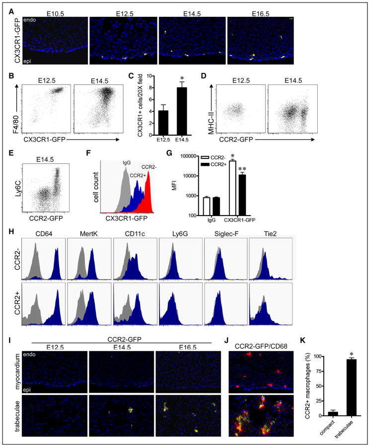
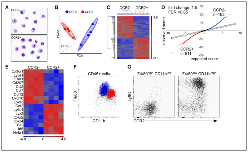
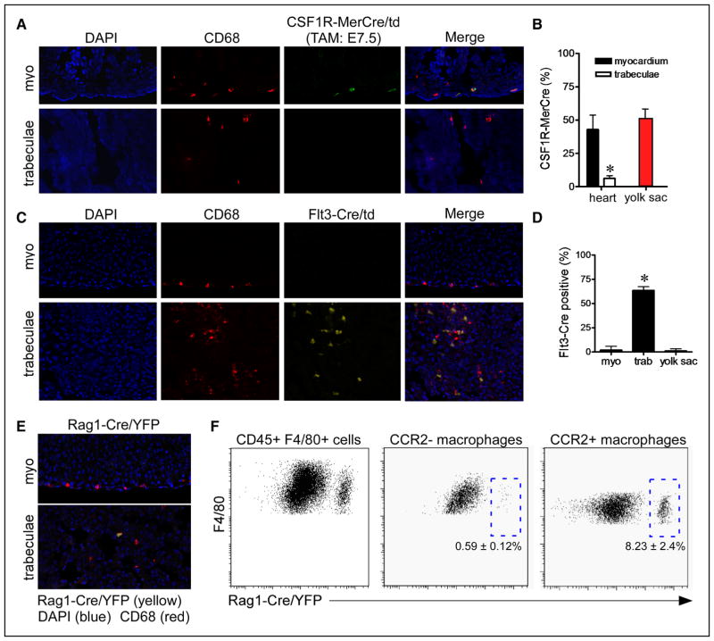
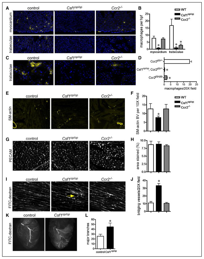
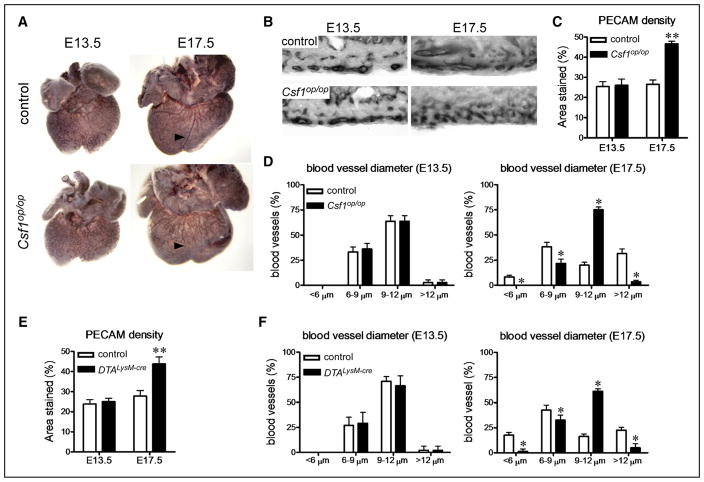
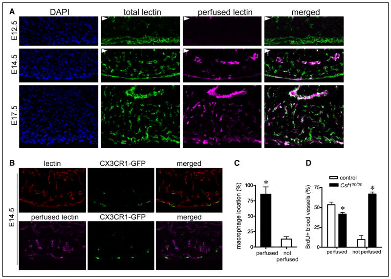
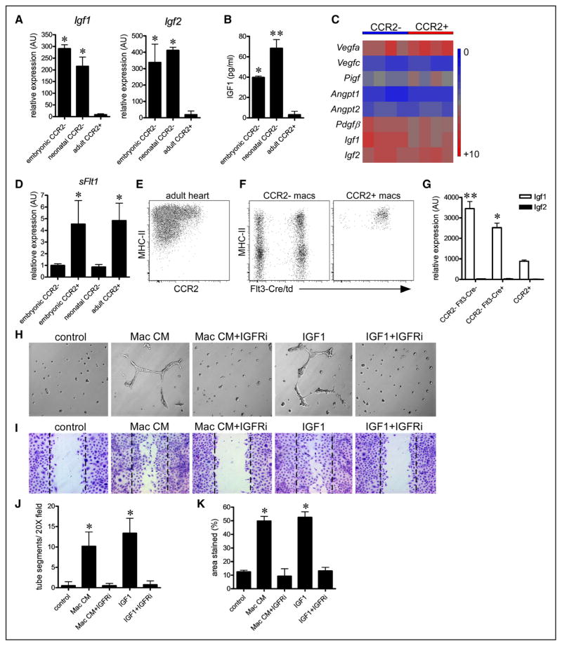
Methods and results: Using a combination of flow cytometry, immunostaining, and genetic lineage tracing, we demonstrate that the developing heart contains a complex array of embryonic macrophage subsets that can be divided into chemokine (C-C motif) receptor 2(-) and chemokine (C-C motif) receptor 2(+) macrophages derived from primitive yolk sac, recombination activating gene 1(+) lymphomyeloid, and Fms-like tyrosine kinase 3(+) fetal monocyte lineages. Functionally, yolk sac-derived chemokine (C-C motif) receptor 2(-) macrophages are instrumental in coronary development where they are required for remodeling of the primitive coronary plexus. Mechanistically, chemokine (C-C motif) receptor 2(-) macrophages are recruited to coronary blood vessels at the onset of coronary perfusion where they mediate coronary plexus remodeling through selective expansion of perfused vasculature. We further demonstrate that insulin like growth factor signaling may mediate the proangiogenic properties of embryonic-derived macrophages.

Conclusions: Together, these findings demonstrate that the embryonic heart contains distinct lineages of embryonic macrophages with unique functions and reveal a novel mechanism that governs coronary development.

Keywords: flow cytometry; fms-like tyrosine kinase 3; macrophages; monocytes; yolk sac.


Figures

Figure 1
Figure 1. Distinct chemokine (C-C motif) receptor 2 (CCR2) and CCR2+ macrophages populate the embryonic heart
A, CX3C chemokine receptor 1 (CX3CR1)-green florescent protein (GFP) embryos demonstrating that macrophages are present in the embryonic heart beginning after E10.5. DAPI (4′,6-diamidino-2-phenylindole), blue; CX3CR1-GFP, yellow; B, Flow cytometry analysis of gated CD45+ cells from the embryonic heart showing that all CD45+ cells express CX3CR1-GFP and F4/80. C, Quantification of the number of CX3CR1-GFP+ macrophages per 20× field at E12.5 and E14.5. *P<0.05. D, Flow cytometry demonstrating that at E12.5 the embryonic heart contains a single CCR2 MHC-IIlow macrophage subset, whereas at E14.5, the embryonic heart contains CCR2 MHC-IIlow and CCR2+ major histocompatibility complex (MHC)-IIlow macrophage subsets. E, CCR2+ macrophage coexpress Ly6C. F and G, Quantification of mean florescent intensity (MFI) demonstrating that CCR2 macrophages express CX3CR1-GFP to a greater extent to that of CCR2+ macrophages. *P<0.05 compared with control, **P<0.05 compared with all other groups. H, CCR2 and CCR2+ macrophages express CD64, MertK, and CD11c. Gray: isotype control, blue: designated antibody. I, Analysis of CCR2-GFP embryos revealing that CCR2+ macrophages are specifically located adjacent to the endocardial trabeculae at E14.5 and E16.5. J and K, Immunostaining for CCR2-GFP (yellow) and CD68 (red) demonstrating that CCR2+ macrophages exclusively reside within the endocardial trabeculae and CCR2 macrophages in the compact myocardium. *P<0.05. A, I, and J: ×20 magnification. Each experiment included at least 4 independent biological replicates. Endo indicates endocardium; and Epi, epicardium.
Figure 2
Figure 2. Gene expression profiling of embryonic cardiac macrophages
A, Cytospin specimens of chemokine (C-C motif) receptor 2 (CCR2) and CCR2+ macrophage subsets sorted by flow cytometry demonstrating robust purification of cell populations. B and C, Principal components analysis (PCA) (B) and hierarchical clustering (C) analyses showing that CCR2 and CCR2+ macrophages have distinct gene expression profiles. D, Significance analysis of microarrays plot highlighting the number of genes upregulated in CCR2 (blue, n=163) and CCR2+ (red, n=511) macrophages. E, Heat map depicting monocyte and macrophage-associated genes that distinguish embryonic CCR2 and CCR2+ macrophages. F and G, Flow cytometry at E14.5 revealing the surface phenotype of CCR2 macrophages (blue): F4/80highCD11blowLy6Cneg and CCR2+ macrophages (red): F4/80lowCD11bhighLy6Cpos. Microarray experiments included 4 biologically independent samples each of which containing 4 to 6 E14.5 hearts. FDR indicates false discovery rate.
Figure 3
Figure 3. Developmental origins of embryonic cardiac macrophages
A, Genetic lineage tracing using colony-stimulating factor 1 receptor (Csf1R)-murine estrogen receptor Cre (MerCre); reverse orientation splice acceptor (Rosa) 26-td (green) demonstrating that tamoxifen administration at E7.5 exclusively labels a subset of CD68+ macrophages (red) located within the compact myocardium and does not label CD68+ macrophages located in the trabeculae. Immunostaining was performed at E14.5. B, Quantification of the percent of total macrophages labeled by Csf1R-MerCre; Rosa26-td reveals that cardiac macrophages located within the myocardium are labeled at a frequency similar to yolk sac macrophages. Five independent hearts were included in the quantitative analysis. C, Fms-like tyrosine kinase (Flt) 3-Cre lineage tracing at E14.5 (yellow) demonstrating exclusive labeling of CD68+ macrophages (red) located in the trabeculae. D, Quantification of CD68 and Flt3-Cre/Rosa26td staining showing that >60% of macrophages located within the trabeculae are Flt3-Cre/Rosa26td positive. In contrast, only rare cells were labeled in the compact myocardium and yolk sac. Five independent hearts were included in the quantitative analysis. E, Recombination activating gene 1 (Rag1)-Cre lineage tracing at E14.5 (yellow) revealing infrequent labeling of CD68+ macrophages (red) located within the trabeculae. F, Flow cytometry analysis of CD45+, CD11b+, F4/80+ cells demonstrating that Rag1-Cre labels <10% of chemokine (C-C motif) receptor 2 (CCR2)+ macrophages. Quantitation is based on 3 independent pools of Rag1-Cre; Rosa26-yellow florescent protein (YFP) embryonic hearts. A, C, and E, ×20 magnification. *P<0.05 compared with all other groups. DAPI indicates 4′,6-diamidino-2-phenylindole.
Figure 4
Figure 4. Embryonic cardiac macrophages are essential for coronary patterning
A and B, CD68 immunostaining (A) and quantification (B) at E16.5 demonstrating that Csf1op/op embryos lack all macrophage subsets, whereas Ccr2−/− embryos specifically have decreased numbers of macrophages within the trabeculae. C and D, chemokine (C-C motif) receptor 2 (CCR2)-green florescent protein (GFP) reporter imaging at E16.5 demonstrating marked reductions in the number of CCR2+ macrophages (yellow) in Csf1op/op and Ccr2−/− hearts. E and F, Smooth muscle (SM) actin immunostaining at P21 showing reduced number of smooth muscle+ arterioles in Csf1op/op hearts. G and H, Platelet endothelial cell adhesion molecule (PECAM) immunostaining at P21 showing unaffected capillary density between genotypes. I, Compressed z-stack images of hearts perfused with fluorescein isothiocyanate (FITC)-dextran demonstrating increased connections (arrow: bridging blood vessels) within the microvasculature. Photographs were obtained from areas of the heart where cardiomyocytes were oriented in parallel. J, Quantification of bridging blood vessels. K and L, FITC dextran perfusion at P21 revealing excessive coronary branching in Csf1op/op mice. Major branches were defined as primary and secondary branches arising from the left anterior descending artery. E, ×10 magnification. A, C, G, and I: ×20 magnification. *P<0.05 compared with controls. Each experiment included at least 5 hearts per genotype. WT indicates wild-type.
Figure 5
Figure 5. Embryonic macrophages function to remodel the developing coronary vascular plexus
A, Platelet endothelial cell adhesion molecule (PECAM) whole-mount immunostaining demonstrating normal coronary plexus development at E13.5 and aberrant coronary patterning at E17.5 in Csf1op/op hearts. Arrowhead denotes expected location of a mature coronary vessel. B, Cryosections of PECAM-stained embryonic hearts demonstrating increased capillary density and failure of the coronary plexus to remodel into blood vessels of varying diameters at E17.5 in Csf1op/op hearts. C and D, Quantification of coronary density (C) and blood vessel diameter (D) in control and Csf1op/op hearts. Csf1op/op hearts have increased coronary density at E17.5 and display failure to remodel the vascular plexus into larger and smaller blood vessels. E and F, PECAM immunostaining demonstrating increased coronary vascular plexus density (E) and defective coronary plexus remodeling (F) in reverse orientation splice acceptor (Rosa) 26-DTALysM-Cre hearts at E17.5. Each experiment included at least 5 hearts per genotype. B, ×20 magnification. *P<0.05 compared with controls.
Figure 6
Figure 6. Chemokine (C-C motif) receptor 2 (CCR2) embryonic macrophages are selectively associated with perfused coronary plexus vasculature
A, Simultaneous imaging of total coronary vasculature (green, fluorescein isothiocyanate-lectin staining) and perfused coronary vasculature (magenta, perfused rhodamine-lectin) demonstrating the onset of coronary perfusion at approximately E14.5. Arrowheads denote the endocardium. B, Perfused (magenta) and total (red) lectin imagining at E14.5 revealing that CX3C chemokine receptor 1 (CX3CR1)-green florescent protein (GFP)+ macrophages are selectively associated with perfused coronary vasculature. C, Quantification of the percent of macrophages localized adjacent to perfused and nonperfused coronary plexus vasculature. D, Bromodeoxyuridine (BrdU) immunostaining showing reduced proliferation of perfused coronary vasculature and markedly increased proliferation of nonperfused vasculature in Csf1op/op hearts. Each experiment included at least 5 hearts. A and B, ×20 magnification. *P<0.05 compared with controls.
Figure 7
Figure 7. Insulin like growth factor (IGF) ligands constitute a proangiogenic cardiac macrophage-derived signal
A, Quantitative reverse transcription polymerase chain reaction (RT-PCR) revealing selective expression of Igf1 and Igf2 mRNA in embryonic chemokine (C-C motif) receptor 2 (CCR2) and neonatal CCR2 macrophages. B, IGF1 ELISA demonstrating that conditioned media obtained from embryonic CCR2 and neonatal CCR2 macrophages contains higher concentrations of IGF1 protein compared with conditioned media obtained from adult CCR2+ macrophages. n=4 for each macrophage population. *P<0.05 compared with adult CCR2+ macrophages. **P<0.05 compared with all other groups. C, Heat map depicting proangiogenic growth factor expression in embryonic CCR2 and CCR2+ macrophages. D, Quantitative RT-PCR demonstrating that embryonic and adult CCR2+ macrophages express higher levels of the angiogenesis inhibitor, soluble Flt1. E and F, Flow cytometry of adult cardiac macrophages revealing the presence of CCR2 and CCR2+ macrophage populations. Lineage tracing shows that CCR2 macrophages can be divided into embryonic-derived (Flt3-Cre negative) and monocyte-derived (Flt3-Cre positive) subsets, whereas CCR2+ macrophages are exclusively monocyte derived. Macrophages were identified as CD45+ CD64+ Ly6G cells. G, Quantitative RT-PCR demonstrating increased expression of Igf1 in CCR2 macrophages compared with CCR2+ macrophages. CCR2 embryonic-derived macrophages express slightly higher levels of Igf1 mRNA compared with CCR2 monocyte-derived macrophages. Igf2 expression could not be detected in adult cardiac macrophages. *P<0.05 to adult CCR2+ macrophages. **P<0.05 compared with all other groups. G, Matrigel angiogenesis assays demonstrating that inhibition of IGF signaling suppresses the ability of CCR2 embryonic macrophage conditioned media to stimulate endothelial cell tube formation in vitro. H, Endothelial scratch assays revealing that inhibition of IGF signaling suppresses the ability of CCR2 embryonic macrophage conditioned media to stimulate endothelial cell migration in vitro. I and J, Quantitative analysis of Matrigel angiogenesis (I) and scratch assays (J). Each experiment included at least 5 biological replicates. H and I, ×20 magnification. *P<0.05 compared with vehicle control.

References

    1. Epelman S, Lavine KJ, Randolph GJ. Origin and functions of tissue macrophages. Immunity. 2014;41:21–35. doi: 10.1016/j.immuni.2014.06.013. - DOI - PMC - PubMed
    1. Davies LC, Jenkins SJ, Allen JE, Taylor PR. Tissue-resident macrophages. Nat Immunol. 2013;14:986–995. doi: 10.1038/ni.2705. - DOI - PMC - PubMed
    1. Ajami B, Bennett JL, Krieger C, McNagny KM, Rossi FM. Infiltrating monocytes trigger EAE progression, but do not contribute to the resident microglia pool. Nat Neurosci. 2011;14:1142–1149. doi: 10.1038/nn.2887. - DOI - PubMed
    1. Boyer SW, Schroeder AV, Smith-Berdan S, Forsberg EC. All hematopoietic cells develop from hematopoietic stem cells through Flk2/Flt3-positive progenitor cells. Cell Stem Cell. 2011;9:64–73. doi: 10.1016/j.stem.2011.04.021. - DOI - PMC - PubMed
    1. Böiers C, Carrelha J, Lutteropp M, et al. Lymphomyeloid contribution of an immune-restricted progenitor emerging prior to definitive hematopoietic stem cells. Cell Stem Cell. 2013;13:535–548. doi: 10.1016/j.stem.2013.08.012. - DOI - PubMed

Publication types

MeSH terms

Substances