. 2016 Mar 24:6:23464.
doi: 10.1038/srep23464.

Hepatitis C virus NS5A protein cooperates with phosphatidylinositol 4-kinase IIIα to induce mitochondrial fragmentation

Affiliations

Hepatitis C virus NS5A protein cooperates with phosphatidylinositol 4-kinase IIIα to induce mitochondrial fragmentation

Gavin Ka Yu Siu et al. Sci Rep. .

Abstract

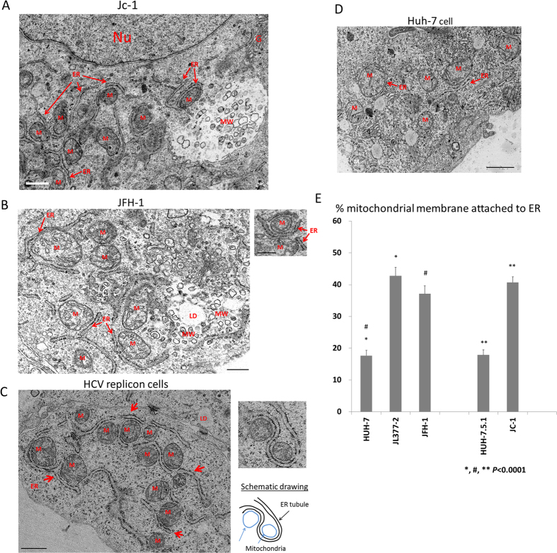
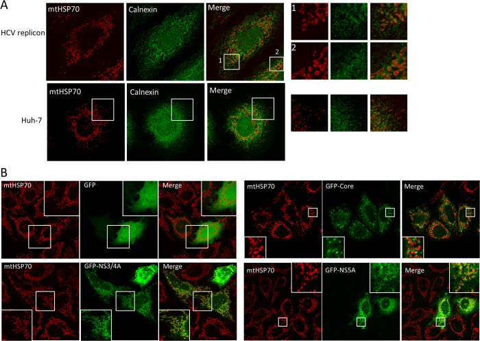
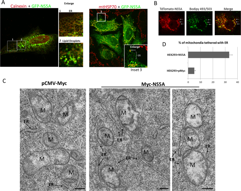
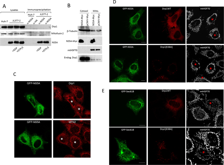
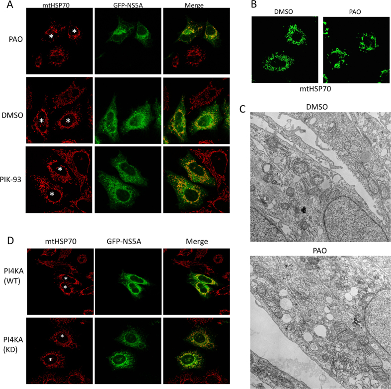
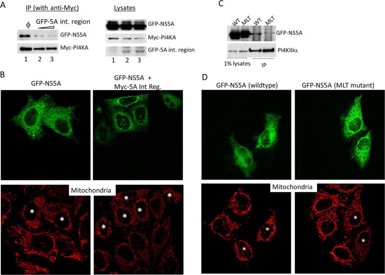
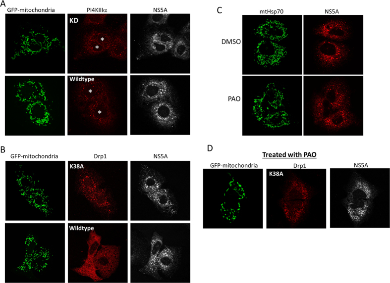
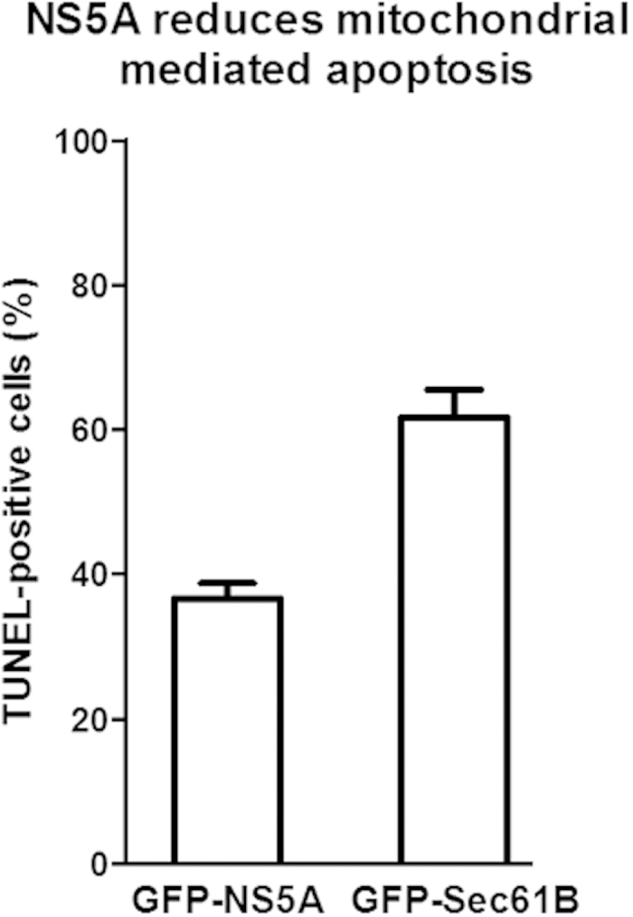
Hepatitis C virus (HCV) has long been observed to take advantage of the host mitochondria to support viral replication and assembly. The HCV core protein has been implicated to fragment host mitochondria. In this report, we have discovered that the non-structural protein 5A (NS5A) plays an instructive role in attaching ER with mitochondria, causing mitochondrial fragmentation. Dynamin-related protein 1(Drp1), a host protein essential to mitochondrial membrane fission, does not play a role in NS5A-induced mitochondrial fragmentation. Instead, phosphatidylinositol 4-kinase IIIα (PI4KA), which has been demonstrated to bind to NS5A and is required to support HCV life cycle, is required for NS5A to induce mitochondrial fragmentation. Both NS5A and core are required by HCV to fragment the mitochondria, as inhibiting either of their respective downstream proteins, PI4KA or Drp1, resulted in lengthening of mitochondria tubules in HCVcc-infected cells. By fragmenting the mitochondria, NS5A renders the cells more resistant to mitochondria mediated apoptosis. This finding indicates previously-ignored contribution of NS5A in HCV-induced mitochondria dysfunction.


Figures

Figure 1
Figure 1. Extensive attachment of ER tubules to mitochondria in HCV replicon cells JL377-2 and hepatocytes transfected with JFH-1 or infected with HCVcc.
(A) Electron micrographs of Huh-7.5.1 cells infected with HCVcc. Scale bar = 0.5 μm. (B) EM images of Huh-7 cells transfected with JFH-1 full-length HCV genomic RNA. Scale bar = 0.5 μm. (C) EM images of the cytoplasm of a JL377-2 cell. Scale bar = 1μm. (D) EM image of naïve Huh-7 cells (left panel). Scale bar = 1μm. Abbreviations: M, mitochondria; ER, endoplasmic reticulum; G, Golgi; Nu, nucleus; MW, membranous web, LD, lipid droplet. (E) Quantification of the amount of mitochondria membrane tethered with ER. The percentage of perimeter membrane of mitochondria closely apposed to ER tubules in EM images of the indicated HCV harboring cells or naïve Huh-7 or Huh-7.5.1 cells were quantified. N = 60; error bars = S.E.M.; p < 0.0001.
Figure 2
Figure 2. NS5A and NS3/4A play a role in tethering ER and mitochondria.
(A) Immunofluorescence images of HCV replicon cells JL-377-2 and naïve Huh-7 cells staining with mitochondria marker mtHSP70 (red) and ER marker Calnexin (green). (B) GFP (top row of panels), GFP-NS3/4A (second row), GFP-core (third row) and GFP-NS5A (bottom row) were expressed in HEK293 cells and tested for their ability to attach to mitochondria (mtHSP70).
Figure 3
Figure 3. NS5A expression in HEK293 cells and Huh-7 cells.
(A) GFP-NS5A is localized to ER membranes (inset 1 and bracket, inset 3) and induces formation of lipid droplets (inset 2). GFP-NS5A frequently fragments and encircles mitochondria (arrow, inset 3). In this experiment, HEK293 cells were transfected with GFP-NS5A for 18 hours before fixation and staining with mtHSP70 (red). (B) TdTomato-NS5A (red) transfected into Huh-7 cells and stained with lipid droplet marker bodipy 493/503 (green). NS5A encircles lipid droplets (arrows). (C) EM study of the effect of NS5A in ER-mitochondria tethering in HEK293 cells transfected with Myc-NS5A or pMyc vector. Extensive ER-tethered mitochondria was observed in Myc-NS5A transfected HEK293 cells but not in Myc vector transfected cells. Scale bars = 1 μm. Abbreviations: M, mitochondria; ER, endoplasmic reticulum. (D) Quantification of ER-mitochondria tethering in NS5A-transfected HEK293 cells from EM images. The percentage of ER-tethered mitochondria is determined by the length of the mitochondria perimeter apposed to ER tubules in the EM images. N = 60; error bar = S.E.M.
Figure 4
Figure 4. NS5A-induced mitochondrial fragmentation is independent of Drp1.
(A) NS5A does not interact with Drp1. Huh-7 or JL377-2 cell lysates were subjected to immunoprecipitation by α-NS5A antibody in the presence of GDP or GTPγS. A monoclonal antibody against HA was used as a control. In all conditions, neither endogenous Drp1 nor mitofusin 2 was co-precipitated with NS5A. (B) NS5A does not increase the mitochondrial association of Drp1. HEK293 cells overexpressed with Myc-NS5A or control Myc-empty vector and the extent of Drp1 on mitochondria-enriched membranes were determined by immunoblotting. (C) Overexpression of GFP-NS5A does not change the subcellular localization of endogenous Drp1 and mitofusin 2 (MFN2) by immunofluorescence. GFP-NS5A expressing cells are marked with asterisks. (D) Wildtype Drp1 or Drp1[K38A] dominant negative mutant cDNA sequences (red) were cotransfected with GFP-NS5A (green) into HEK293 cells. The mitochondria were visualized by staining the cells with mtHSP70 at 633 nm wavelength excitation laser (white). (E) Drp1 or Drp1[K38A] dominant negative mutant cDNA sequences (red) were cotransfected with control GFP-Sec61B (green) into HEK293 cells. The mitochondria were visualized by staining the cells with mtHSP70 at 633 nm wavelength excitation laser (white). Red asterisks indicate transfected cells. Scale Bar = 10 μm.
Figure 5
Figure 5. PI4KA is required for NS5A to cause mitochondrial fragmentation.
(A) PI4KA inhibitor PAO but not PI4KB inhibitor PIK-93 or vehicle DMSO modulates the effect of NS5A on mitochondrial fragmentation. HEK293 cells transfected with GFP-NS5A were treated with the indicated inhibitors before immunofluorescence staining. Asterisks indicate the mitochondria of GFP-NS5A transfected cells. (B) JL377-2 HCV replicon cells treated with PAO were stained with mitochondria marker mtHSP70. (C) EM ultrastructures of JL377-2 cells treated with DMSO or PAO. (D) Co-expression of the kinase deficient mutant of PI4KA modulates the mitochondrial fragmentation caused by GFP-NS5A in HEK293 cells. GFP-NS5A and PI4KA DNA constructs were transfected at ratio of 1:5 into HEK293 cells. The cells were subjected to immunofluorescence staining using antibody against mtHSP70 to label the mitochondria (red). Asterisks mark the cells expressing the indicating DNA constructs.
Figure 6
Figure 6. Interaction between NS5A and PI4KA is critical for the NS5A-induced mitochondrial fragmentation.
(A) Disruption of the NS5A-PI4KA interaction by overexpression the NS5A-interacting region of PI4KA in HEK293T cells. The protein interaction between GFP-NS5A and Myc-PI4KA was investigated by immunoblotting after immunoprecipitation using anti-Myc antibody. The interaction between GFP-NS5A and Myc-PI4KA can be disrupted by co-expression of GFP-5A int. region (lanes 2 and 3). (B) Disruption of NS5A-PI4KA interaction can abrogate the mitochondrial fragmentation activity of NS5A. GFP-NS5A alone or in combination with Myc-5A interacting region (Myc-5A Int Reg.) at a ratio of 1:5 was transfected into HEK293 cells which were subsequently stained with mitochondria marker mtHSP70. Transfected cells are indicated with asterisks. (C) NS5A with mutations at the PI4KA binding motif (MLT mutant) shows significantly reduced binding to PI4KA. GFP-NS5A wildtype (WT) or MLT mutant (MLT) were co-expressed with Myc-PI4KA in HEK293T cells and immunoprecipitated with anti-Myc 9E10 antibody (bottom panel). The extent of co-precipitated GFP-NS5A proteins was investigated by immunoblotting (top panel). (D) Overexpression of the NS5A MLT mutant no longer caused mitochondrial fragmentation. GFP-NS5A wildtype or MLT mutant were expressed in HEK293 cells (top panels) and the status of mitochondria was analyzed with immunofluorescence staining of mtHSP70 (bottom panels). Asterisks indicate the corresponding NS5A transfected cells.
Figure 7
Figure 7. Drp1 and PI4KA are both necessary in mitochondrial fragmentation in HCV infected cells.
(A) Mitochondrial fragmentation is reduced by the kinase deficient (KD) mutant of PI4KA in Huh-7.5.1 cells infected with HCVcc (Jc-1). Asterisks indicate PI4KA transfected cells. NS5A staining indicates HCV infected cells. (B) Mitochondrial fragmentation is reduced by dominant negative Drp1 mutant (K38A) in HCVcc-infected cells. (C) PI4KA inhibitor PAO reduces mitochondrial fragmentation in Huh-7.5 cells infected with HCVcc. (D) Treating HCVcc infected cells that have been transfected with Drp1 (K38A) with PAO did not cause drastic increase in mitochondria tubular length.
Figure 8
Figure 8. NS5A reduces mitochondrial mediated apoptosis.
GFP-NS5A or control GFP-Sec61B transfected HEK293 cells were treated with hydrogen peroxide before TUNEL staining for detection of apoptotic cells. In five independent experiments, a total of over 500 transfected cells were counted in each condition; error bars = S.E.M.; P < 0.0001.

References

    1. Tai A. W. et al. A Functional Genomic Screen Identifies Cellular Cofactors of Hepatitis C Virus Replication. Cell Host & Microbe 5, 298–307 (2009). - PMC - PubMed
    1. Berger K. L. et al. Roles for endocytic trafficking and phosphatidylinositol 4-kinase III alpha in hepatitis C virus replication. Proceedings of the National Academy of Sciences 106, 7577–7582 (2009). - PMC - PubMed
    1. Reiss S. et al. Recruitment and Activation of a Lipid Kinase by Hepatitis C Virus NS5A Is Essential for Integrity of the Membranous Replication Compartment. Cell Host & Microbe 9, 32–45 (2011). - PMC - PubMed
    1. Berger K. L., Kelly S. M., Jordan T. X., Tartell M. A. & Randall G. Hepatitis C Virus Stimulates the Phosphatidylinositol 4-Kinase III Alpha-Dependent Phosphatidylinositol 4-Phosphate Production That Is Essential for Its Replication. Journal of Virology 85, 8870–8883 (2011). - PMC - PubMed
    1. Lim Y.-S. & Hwang S. B. Hepatitis C Virus NS5A Protein Interacts with Phosphatidylinositol 4-Kinase Type IIIα and Regulates Viral Propagation. Journal of Biological Chemistry 286, 11290–11298 (2011). - PMC - PubMed

Publication types

MeSH terms

Substances