. 2016 Mar 24;7(3):e2151.
doi: 10.1038/cddis.2016.56.

Inhibition of glycogen synthase kinase 3β promotes autophagy to protect mice from acute liver failure mediated by peroxisome proliferator-activated receptor α

Affiliations

Inhibition of glycogen synthase kinase 3β promotes autophagy to protect mice from acute liver failure mediated by peroxisome proliferator-activated receptor α

F Ren et al. Cell Death Dis. .

Abstract

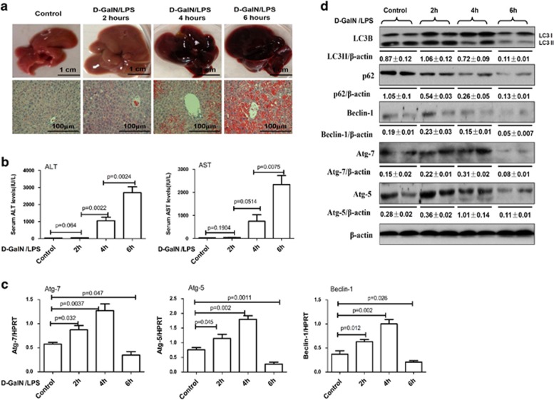
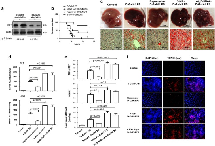
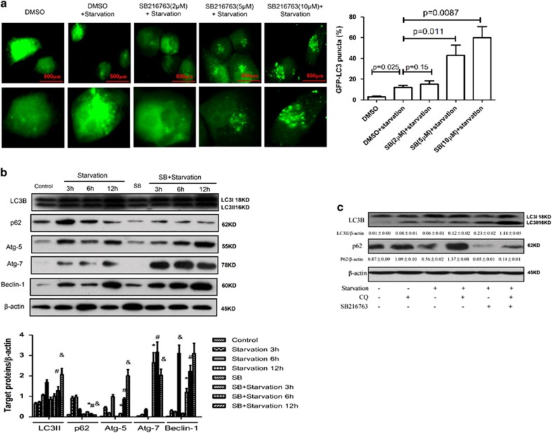
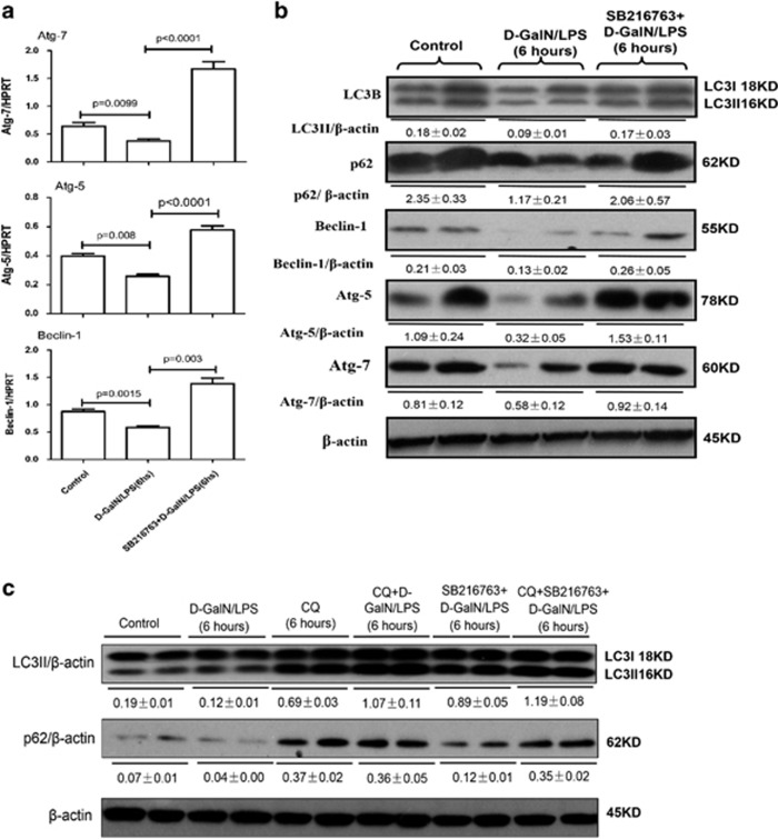
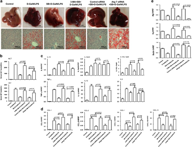
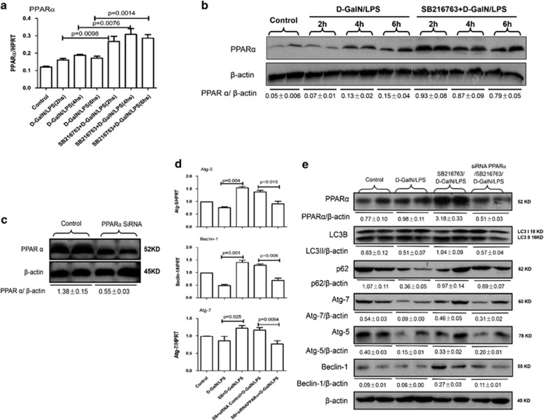
Our previous studies have demonstrated that inhibition of glycogen synthase kinase 3β (GSK3β) activity protects mice from acute liver failure (ALF), whereas its protective and regulatory mechanism remains elusive. Autophagy is a recently recognized rudimentary cellular response to inflammation and injury. The aim of the present study was to test the hypothesis that inhibition of GSK3β mediates autophagy to inhibit liver inflammation and protect against ALF. In ALF mice model induced by D-galactosamine (D-GalN) and lipopolysaccharide (LPS), autophagy was repressed compared with normal control, and D-GalN/LPS can directly induce autophagic flux in the progression of ALF mice. Autophagy activation by rapamycin protected against liver injury and its inhibition by 3-methyladenine (3-MA) or autophagy gene 7 (Atg7) small interfering RNA (siRNA) exacerbated liver injury. The protective effect of GSK3β inhibition on ALF mice model depending on the induction of autophagy, because that inhibition of GSK3β promoted autophagy in vitro and in vivo, and inhibition of autophagy reversed liver protection and inflammation of GSK3β inhibition. Furthermore, inhibition of GSK3β increased the expression of peroxisome proliferator-activated receptor α (PPARα), and the downregulated PPARα by siRNA decreased autophagy induced by GSK3β inhibition. More importantly, the expressions of autophagy-related gene and PPARα are significantly downregulated and the activity of GSK3β is significantly upregulated in liver of ALF patients with hepatitis B virus. Thus, we have demonstrated the new pathological mechanism of ALF that the increased GSK3β activity suppresses autophagy to promote the occurrence and development of ALF by inhibiting PPARα pathway.


Figures

Figure 1
Figure 1
The regulation of autophagy during d-GalN/LPS induced mice ALF progression. Mice were intraperitoneally injected with d-GalN (700 mg/kg) and LPS (10 μg/kg) at 2, 4 and 6 h (12 mice per group). The mice in the control group (n=8) were injected with PBS only. (a). Representative livers and H&E staining of livers. (b) Serum AST and ALT enzyme levels. (c) Gene expressions of Beclin-1, Atg5 and Atg7 were measured by qRT-PCR in the livers. (d) Protein expression levels of LC3B, p62, Beclin-1, Atg5 and Atg7 were measured by western blot assays in the livers. A representative blot from two samples of every group is shown
Figure 2
Figure 2
Autophagy protects against d-GalN/LPS-induced liver injury rapamycin+d-GalN/LPS-treated mice were administered rapamycin (2 mg/kg, i.p.) 2 h before d-GalN/LPS exposure (n=12); 3-MA+d-GalN/LPS-treated mice were administered 3-MA (10 mg/kg, i.p.) 2 h before d-GalN/LPS exposure (n=12); siRNA Atg7+d-GalN/LPS-treated mice were pretreated with siRNA Atg7 (3 mg/kg, i.v.) for 48 h before d-GalN/LPS exposure (n=12). Control mice were pretreated with vehicle (DMSO) 2 h before PBS injection (n=8). (a) Protein expression levels of Atg7 and β-actin were measured by western blotting. A representative blot for two samples from every group is shown. (b) The survival rate of mice was measured in the Atg7 siRNA +d-GalN/LPS-, rapamycin+d-GalN/LPS- and 3-MA+d-GalN/LPS-treated group and the d-GalN/LPS-treated group (10 mice per group). (c) Representative livers and H&E staining of livers from different groups. (d) Serum AST and ALT enzyme levels from different groups. (e) Gene expression levels of TNF-α and IL-6 were measured by qRT-PCR and the level of MDA was measured in livers from different groups. (f) TUNEL staining (red) liver tissue at 6 h after d-GalN/LPS administration. Representative of one experiment is shown. Original magnification × 200
Figure 3
Figure 3
Inhibition of GSK3β further promotes autophagy induced by starvation in vitro. (a). Transfected GFP-LC3 plasmid for 12 h, the primary hepatocytes were pre-incubated with SB216763 (2, 5 or 10 μM) for 12 h to observe the formation of autophagosomes. (b) The primary hepatocytes were stimulated with or without SB216763 for 30 min, followed for 3, 6 or 12 h. The cell lysates were analyzed by western blot in different time. The levels of LC3B, p62, Atg7, Atg5, Beclin-1and β-actin were measured by western blotting. The graph of densitometry analysis should be shown (compared with starvation 3 h group: *P<0.05; compared with starvation 6 h group: #P<0.05; compared with starvation 12 h group: &P<0.05). (c) Inhibition of GSK3β induced autophagic flux in primary hepatocytes under starvation. Western blot analysis of expression of the LC3B and p62. A representative results from three independent experiments is shown. Densitometry analysis of the proteins was performed for each sample
Figure 4
Figure 4
Autophagy is promoted by GSK3β signaling molecule in vivo. The mice were pretreated with or without SB216763 (25 mg/kg, i.p.) for 2 h, followed by d-GalN/LPS for 6 h stimulation (n=10). CQ-treated mice were pretreated with CQ (60 mg/kg, i.p.) for 8 h (n=10); CQ+ d-GalN/LPS-treated mice were pretreated with CQ (60 mg/kg, i.p.) for 2 h before d-GalN/LPS exposure (n=10); CQ+SB216763+d-GalN/LPS-treated mice were pretreated with CQ (60 mg/kg, i.p.) and SB216763 for 2 h before d-GalN/LPS exposure (n=10). (a) Gene expression levels of autophagy-related proteins, including Atg7, Atg5 and Beclin-1 were measured by qRT-PCR in livers of control mice group, d-GalN/LPS-induced ALF mice group and SB216763 pretreament ALF group induced by d-GalN/LPS. (b) Protein expression levels of autophagy-related proteins, including LC3B, p62, Atg7, Atg5 and Beclin-1, were measured by western blotting in livers. Two representative blot for liver samples from each group were shown. (c) Protein expression levels of LC3B and p62 were measured by western blotting in livers. A representative blot for three samples from each group is shown
Figure 5
Figure 5
The inhibition of GSK3β activity protects mice from ALF through autophagy mechanisms. The mice were pretreated with or without SB216763 (25 mg/kg, i.p.) for 2 h, followed by d-GalN/LPS for 6 h stimulation (n=10). SB216763+3-MA+ d-GalN/LPS-treated mice were co-administered 3-MA (10 mg/kg) and SB216763 at 2 h before d-GalN/LPS exposure (n=10); SB216763+siAtg7+ d-GalN/LPS-treated mice were pretreated with Atg7 siRNA (3 mg/kg) for 48 h via tail vein injection and then administered SB216763 2 h before d-GalN/LPS exposure (n=11). (a) Representative livers and H&E staining of livers (200 ×) from different groups. (b) Serum AST and ALT enzyme levels from different groups. (c) Gene expression of cytokines including TNF-α, IL-1β and IL-6 at 6 h, IL-12p40 and IL-10 at 2 h after d-GalN/LPS injection. (d) Gene expression of chemokines including CCL-1, CCL-2, CXCL-1 and CXCL-10 at 6 h after d-GalN/LPS injection. (e) Gene expression of Atg7, Atg5 and Beclin-1 at 6 h after d-GalN/LPS injection
Figure 6
Figure 6
Autophagy is regulated by GSK3β-PPARα signaling pathway in ALF. (a and b) The mice were pretreated with or without SB216763 (25 mg/kg, i.p.) for 2 h, followed by d-GalN/LPS for 2, 4 or 6 h stimulation(n=10 mice per every group). Gene and protein expression levels of PPARα at 2, 4 and 6 h after d-GalN/LPS injection in livers from every groups. A representative blot for two samples from each group is shown. (c) SiRNA control- or siRNA PPARα-treated mice were injected with SiRNA control or PPARα siRNA (3 mg/kg) for 48 h via tail vein injection (n=10); Mice were sacrificed 48 h after siRNA PPARα treatment. Protein expression level of PPARα was measured by western blotting in livers. A representative blot for two samples from each group is shown. (d and e) SB+siRNA PPARα+d-GalN/LPS-treated mice were pretreated with PPARα siRNA (3 mg/kg) for 48 h via tail vein injection and then administered SB216763 2 h before d-GalN/LPS exposure (n=12); SB+siRNA control+d-GalN/LPS-treated mice were pretreated with control siRNA (3 mg/kg) for 48 h via tail vein injection, then administered SB216763 2 h before d-GalN/LPS exposure (n=10). Gene expression levels of autophagy-related proteins were measured by qRT-PCR in livers. Protein expression levels of PPARα and autophagy-related proteins were measured by western blotting in livers. Two representative blot for liver samples from each group were shown
Figure 7
Figure 7
Suppression of autophagy in the liver of ALF patients with HBV infection. (a) Gene expression levels of Atg7, Atg5 and Beclin-1 were measured by qRT-PCR in the livers of normal subjects (n=9), CHB patients (n=14) and ALF patients (n=19). The average target gene/HPRT ratios for each experimental group were plotted. (b) Protein expression levels of LC3B, p62, Atg7, Atg5 and Beclin-1 were measured by western blotting in the livers of normal subjects (n=9), CHB patients (n=14) and ALF patients (n=19). A representative blot from three samples of every group is shown. (c) Protein expression levels of p-GSK3β, GSK3β and PPARα were measured by western blotting in the livers of normal subjects (n=9), CHB patients (n=14) and ALF patients (n=19). A representative blot from three samples of every group is shown. (d) In the d-GalN/LPS-induced ALF mice, d-GalN/LPS treatment induces increase of GSK3β activity, and then suppresses the expression level of PPARα, which promotes down-regulation of autophagy and increases the expression of pro-inflammatory cytokines and chemokines. These events lead to incremental liver inflammation, and promote the hepatocyte apoptosis and ultimately induce the development of ALF

References

    1. Rolando N, Wade J, Davalos M, Wendon J, Philpott-Howard J, Williams R. The systemic inflammatory response syndrome in acute liver failure. Hepatology 2000; 32: 734–739. - PubMed
    1. Hoofnagle JH, Carithers RJ, Shapiro C, Ascher N. Fulminant hepatic failure: summary of a workshop. Hepatology 1995; 21: 240–252. - PubMed
    1. Cohen P, Goedert M. GSK3 inhibitors: development and therapeutic potential. Nat Rev Drug Discov 2004; 3: 479–487. - PubMed
    1. Rayasam GV, Tulasi VK, Sodhi R, Davis JA, Ray A. Glycogen synthase kinase 3: more than a namesake. Br J Pharmacol 2009; 156: 885–898. - PMC - PubMed
    1. Martin M, Rehani K, Jope RS, Michalek SM. Toll-like receptor-mediated cytokine production is differentially regulated by glycogen synthase kinase 3. Nat Immunol 2005; 6: 777–784. - PMC - PubMed

Publication types

MeSH terms

LinkOut - more resources