

Review
doi: 10.1093/jhps/hnu011.
eCollection 2014 Oct.
The role of hip arthroscopy in the management of osteonecrosis
Affiliations
- PMID: 27011804
- PMCID: PMC4765287
- DOI: 10.1093/jhps/hnu011
Item in Clipboard
Review
The role of hip arthroscopy in the management of osteonecrosis
J Hip Preserv Surg.
.
Abstract
Hip arthroscopy has emerged as a diagnostic and therapeutic tool in the management of osteonecrosis (ON) of the femoral head. Direct visualization of the joint, aids the staging of the disease, while mechanical symptoms and pain can be alleviated by addressing the often coexisting intra-articular pathology (labral tears, chondral delamination, loose bodies and synovitis) thereby improving the clinical outcome in some patients. The article explores the role and possible value of hip arthroscopy as a surgical technique in the treatment of hip ON.
Figures
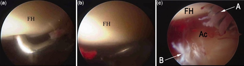
(a) Arthroscopic appearance of a positive ballottement test where the articular cartilage of the femoral head buckles under the pressure of the probe. (b) The articular cartilage of the femoral head assumes its original shape after the release of pressure. (c) In the same hip, associated synovitis and a degenerative labral tear is seen. FH, femoral head; Ac, Acetabulum; a, synovitis, b labrum degeneration).
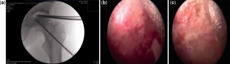
(a) During bone endoscopy the −70 camera is positioned in the core track in order to ensure, that this is positioned directly to the necrotic area. (b) The camera reveals the healthy area of the femoral head as red-bleeding bone.(c) The camera in the necrotic area of the femoral head reveals sclerotic-white bone. (The metal appearing at the top end of the picture is a thin needle that washes out the track to improve our imaging capability).
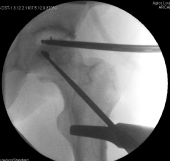
Arthroscopic assisted core decompression and curettage of the necrotic lesion.
References
-
- Lieberman JR, Berry DJ, Mont MA, et al. Osteonecrosis of the hip: management in the 21st century. Instr Course Lect 2003; 52: 337–55. - PubMed
-
- Mont MA, Marulanda GA, Jones LC, et al. Systematic analysis of classification systems for osteonecrosis of the femoral head. J Bone Joint Surg Am 2006; 88(Suppl. 3): 16–26. - PubMed
-
- Malizos KN, Karantanas AH, Varitimidis SE, et al. Osteonecrosis of the femoral head: etiology, imaging and treatment. Eur J Radiol 2007; 63: 16–28. - PubMed
-
- Mont MA, Hungerford DS. Non-traumatic avascular necrosis of the femoral head. J Bone Joint Surg Am 1995; 77: 459–74. - PubMed
-
- National Joint Registry for England and Wales. Annual Clinical Report 2013. Available at: http://www.njrcentre.org.uk. Accessed 24 May 2014.
Publication types
LinkOut - more resources
Full Text Sources
Other Literature Sources
Miscellaneous
