. 2015 Jul;2(2):99-107.
doi: 10.1093/jhps/hnv029. Epub 2015 Jun 6.

Deep gluteal syndrome

Affiliations

Deep gluteal syndrome

Hal David Martin et al. J Hip Preserv Surg. 2015 Jul.

Abstract

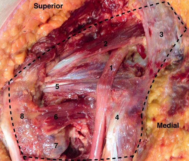
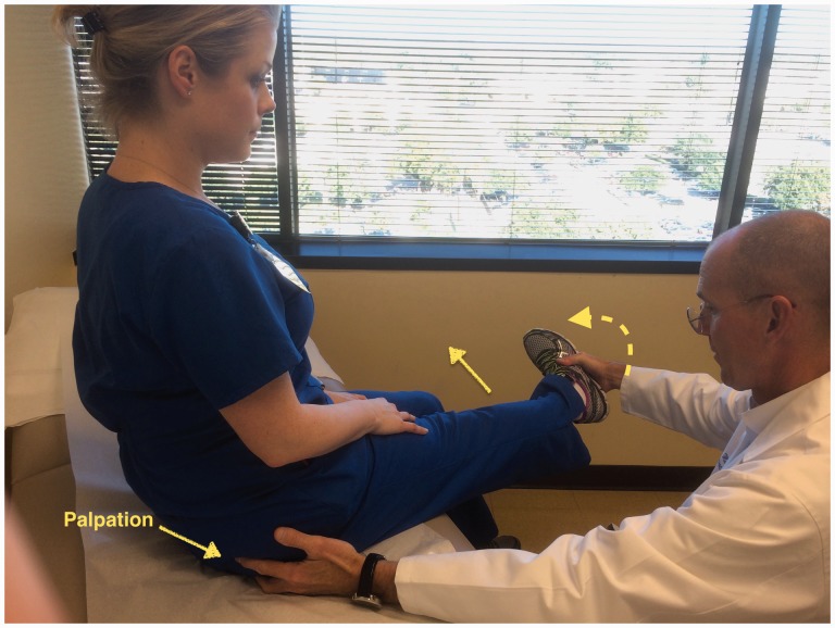
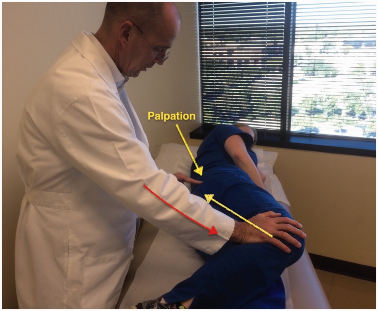
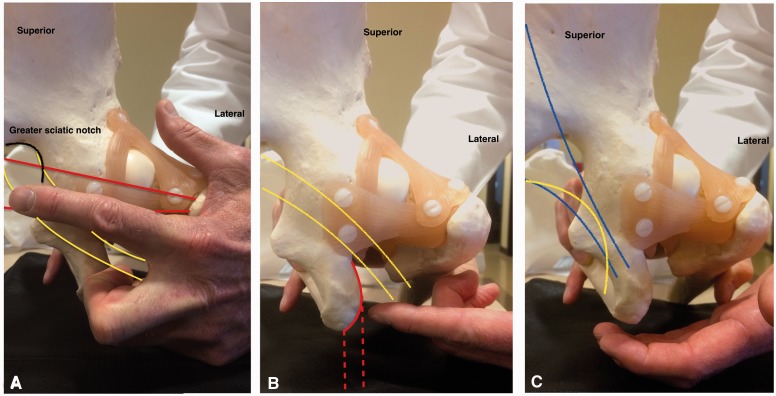
Deep gluteal syndrome describes the presence of pain in the buttock caused from non-discogenic and extrapelvic entrapment of the sciatic nerve. Several structures can be involved in sciatic nerve entrapment within the gluteal space. A comprehensive history and physical examination can orientate the specific site where the sciatic nerve is entrapped, as well as several radiological signs that support the suspected diagnosis. Failure to identify the cause of pain in a timely manner can increase pain perception, and affect mental control, patient hope and consequently quality of life. This review presents a comprehensive approach to the patient with deep gluteal syndrome in order to improve the understanding of posterior hip anatomy, nerve kinematics, clinical manifestations, imaging findings, differential diagnosis and treatment considerations.


Figures

Fig. 1.
Fig. 1.
Deep gluteal space. A cadaveric left hip with the gluteus maximus reflected. The course of the sciatic nerve (1) as it enters the pelvis at the sciatic notch anterior to the piriformis muscle (2) and sacrotuberous ligament (3). As the nerve courses distally toward the ischium and hamstring origin (4) it passes posterior to the gemelli-obturator internus complex (5) and quadratrus femoris (6, with the inferior portion removed to expose the lesser trochanter). Lateral structures include the lesser trochanter (7) and greater trochanter (8).
Fig. 2.
Fig. 2.
Seated piriformis stretch test. The patient is in the seated position, which offers a stable reproducible platform with 90 degrees of hip flexion. The examiner extends the knee (engaging the sciatic nerve) and passively moves the flexed hip into adduction (solid arrow) with internal rotation (dashed arrow) while palpating 1 cm lateral to the ischium (middle finger) and proximally at the sciatic notch (index finger). A positive test is the recreation of the posterior pain at the level of the piriformis or external rotators.
Fig. 3.
Fig. 3.
Active piriformis test. In the lateral position, the patient pushes the heel down into the table and actively abducts with external rotation (yellow arrow) against resistance (orange arrow). The examiner palpates at the level of the piriformis.
Fig. 4.
Fig. 4.
Palpation of the deep gluteal space. (A) At the greater sciatic notch (outlined in black). The path of the piriformis muscle (represented in red) and the sciatic nerve (represented in yellow). (B) Lateral to the ischium (solid red line). The path of the hamstring (dashed red line) and sciatic nerve (represented in yellow). (C) Medial to the ischium. The path of the sacrotuberous ligament (represented in blue) and the pudendal nerve (yellow line).
Fig. 5.
Fig. 5.
Ischiofemoral impingement test. (A) The symptomatic hip is passively taken into extension with zero abduction or adduction. A positive test is the recreation of the posterior hip pain. (B) The symptomatic hip is passively taken into extension with abduction without pain.
Fig. 6.
Fig. 6.
Active hamstring test. (A) The patient performs an active knee flexion against resistance with the knee at 90 degrees. Normal strength without pain may be observed. (B) The patient performs an active knee flexion against resistance with the knee at 30 degrees. Weakness and recreation of the symptoms in this position is a positive test.
Fig. 7.
Fig. 7.
Patient in lateral position, right hip. Observe the location of the anterolateral (AL) portal, posterolateral (PL) portal and the auxiliary posterolateral (APL) portal around the greater trochanter (GT, curved line).
Fig. 8.
Fig. 8.
Sciatic nerve inspection. (A) Normal sciatic nerve appearance with presence of blood flow and epineural fat. (B) Abnormal sciatic nerve with white shoestring appearance, and no epineural fat.
Fig. 9.
Fig. 9.
Passive hip circumductions beggining 45 degrees of hip flexion, maximum external rotation engaging the greater trochanter agains the ischium to mobilize the sciatic nerve lateral.
Fig. 10.
Fig. 10.
Piriformis stretch—in seated position, the patient crosses the leg that will be stretched with the foot positioned next to the knee. The stretching is performed with the patient bringing the knee towards to the contralateral shoulder. The duration and quantity of stretch is determined according with the advance of the healing process.
Fig. 11.
Fig. 11.
Nerve glides—patient in a siting position, hold with both hands under the knee. The exercise is performed with the mobilization of the posterior neural chain. (A) Cervical extension, knee flexion and dorsiflexion. (B) Cervical flexion, knee extension (under the limit for each phase), plantar flexion. The patient can apply lumbar flexion and extension during the exercise.

References

    1. McCrory P, Bell S. Nerve entrapment syndromes as a cause of pain in the hip, groin and buttock. Sports Med 1999; 27: 261–74. - PubMed
    1. Robinson DR. Pyriformis syndrome in relation to sciatic pain. Am J Surg 1947; 73: 355–8. - PubMed
    1. Filler AG, Haynes J, Jordan SE, et al. Sciatica of nondisc origin and piriformis syndrome: diagnosis by magnetic resonance neurography and interventional magnetic resonance imaging with outcome study of resulting treatment. J Neurosurg Spine 2005; 2: 99–115. - PubMed
    1. Martin HD, Shears SA, Johnson JC, et al. The endoscopic treatment of sciatic nerve entrapment/deep gluteal syndrome. Arthroscopy 2011; 27: 172–81. - PubMed
    1. Puranen J, Orava S. The hamstring syndrome: a new diagnosis of gluteal sciatic pain. Am J Sports Med 1988; 16: 517–21. - PubMed