

Preoperative Diagnosis of Juxtaglomerular Cell Tumors in Eight Patients
- PMID: 27012170
- PMCID: PMC8031485
- DOI: 10.1111/jch.12810
Preoperative Diagnosis of Juxtaglomerular Cell Tumors in Eight Patients
Abstract
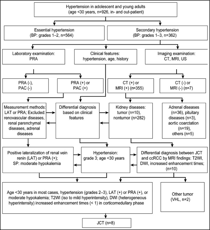
The aim of this study was to improve the diagnostic efficiency for juxtaglomerular cell tumors (JCTs) and to determine whether clinical and magnetic resonance imaging features can help to differentiate JCTs from clear cell renal cell carcinoma (ccRCC). The clinical features of eight patients with JCTs and 27 patients with ccRCCs were analyzed. A flow diagram for young people with hypertension was applied to facilitate the diagnosis. Clinical presentations were analyzed, including age, hypertension, and hypokalemia. The results of our study produced a flow diagram that narrowed the scope of diagnosis. The statistical results demonstrated that patients with a renal mass aged 14 to 30 years, had grade 3 hypertension, or had moderate hypokalemia had a greater possibility of having a JCT than a ccRCC (P<.0000, P<.01, P<.0005, respectively). In addition, the flow diagram and magnetic resonance imaging features were useful to distinguish JCTs from other renal tumors.
©2016 Wiley Periodicals, Inc.
Figures
References
-
- Robertson PW, Klidjian A, Harding LK, et al. Hypertension due to a renin‐secreting renal tumour. Am J Med. 1967;43:963–976. - PubMed
-
- Ørjavik OS, Fauchald P, Hovig T, et al. Renin‐secreting renal tumour with severe hypertension. Case report with tumour renin analysis, histopathological and ultrastructural studies. Acta Med Scand. 1975;197:329–335. - PubMed
-
- Conn JW, Cohen EL, McDonald WJ, et al. Hypertension, hyperreninemia and secondary aldosteronism due to rennin producing juxtaglomerular cell tumor. Arch Intern Med. 1972;13:682–696. - PubMed
-
- McVicar M, Carman C, Chandra M, et al. Hypertension secondary to renin‐secreting juxtaglomerular cell tumor: case report and review of 38 cases. Pediatr Nephrol. 1993;7:404–412. - PubMed
-
- Osawa S, Hosokawa Y, Soda T, et al. Juxtaglomerular cell tumor that was preoperatively diagnosed using selective renal venous sampling. Intern Med. 2013;52:1937–1942. - PubMed
MeSH terms
LinkOut - more resources
Full Text Sources
Other Literature Sources
Medical
