. 2016 Jun 14:325:30-8.
doi: 10.1016/j.neuroscience.2016.03.044. Epub 2016 Mar 21.

Tau pathology-mediated presynaptic dysfunction

Affiliations

Tau pathology-mediated presynaptic dysfunction

H Moreno et al. Neuroscience. .

Abstract

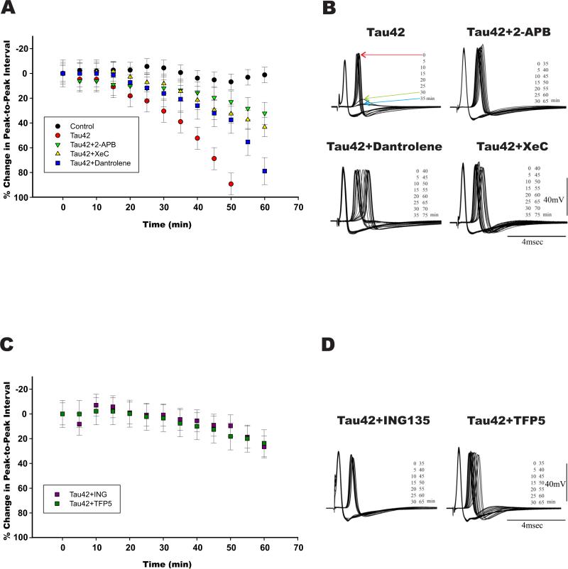
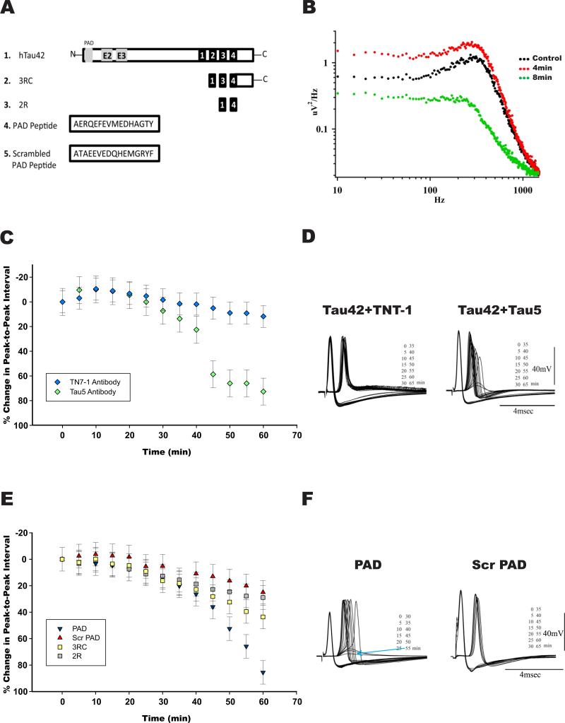
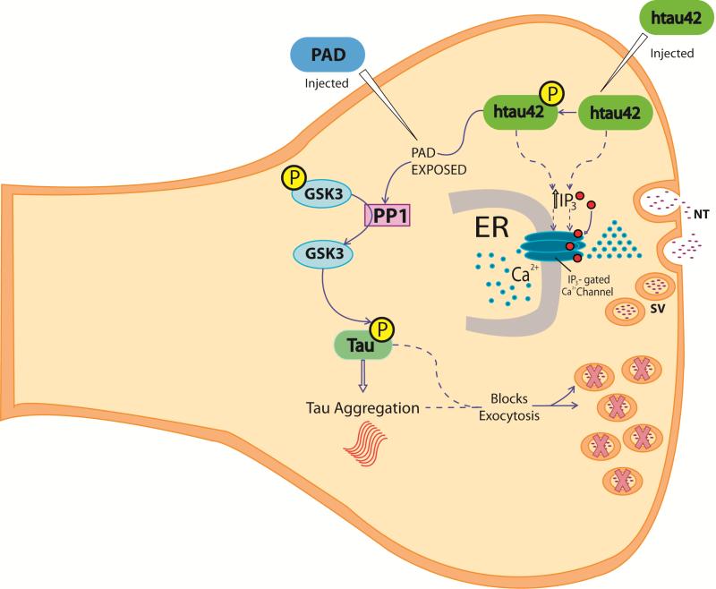
Brain tauopathies are characterized by abnormal processing of tau protein. While somatodendritic tau mislocalization has attracted considerable attention in tauopathies, the role of tau pathology in axonal transport, connectivity and related dysfunctions remains obscure. We have previously shown using the squid giant synapse that presynaptic microinjection of recombinant human tau protein (htau42) results in failure of synaptic transmission. Here, we evaluated molecular mechanisms mediating this effect. Thus, the initial event, observed after htau42 presynaptic injection, was an increase in transmitter release. This event was mediated by calcium release from intracellular stores and was followed by a reduction in evoked transmitter release. The effect of htau42 on synaptic transmission was recapitulated by a peptide comprising the phosphatase-activating domain of tau, suggesting activation of phosphotransferases. Accordingly, findings indicated that htau42-mediated toxicity involves the activities of both GSK3 and Cdk5 kinases.

Keywords: Cdk5; GSK3; IP3 receptor; phosphatase-activating domain of tau; ryanodine receptor; tauopathy.


Figures

Figure 1
Figure 1. Exogenously injected htau42 requires IP3 receptors, GSK3 and Cdk5 activities to induce blockade of synaptic transmission
A) Synaptic transmission time courses: i) vehicle injected axons (black dots). Note that interspike time is maintained relatively constant during the recording period, ii) htau42 injected synapses (red dots), iii) synapses coinjected with: htau42 and 2-APB (green triangles), htau42 and Xestospongin C (yellow triangles) and htau42 and dantrolene (blue squares). Note that synaptic transmission block observed with htau42 was prevented by the IP3Rs inhibitors and delayed by dantrolene. B) Representative pre- and post-synaptic potentials following direct presynaptic axon stimulation every five min, first evoked spike identified by red arrow. Synaptic transmission starts to fail at 30 min, in this example (green arrow), and is completely blocked at 35 min (blue arrow) following h-tau42 preinjection (Tau42; left upper panel). Coinjection of recombinant htau42 protein with the IP3Rs blockers (Tau42+2-APB or Tau42+XeC) prevented htau42 synapto-toxicity for the time of the experiment (65 or 75 min respectively). Dantrolene had a variable response on htau42 toxicity and in this case delayed synaptic block. C) Synaptic transmission time courses for synapses coinjected with htau42 and the GSK3 inhibitor ING-135 (ING, purple squares dots), or with the Cdk5/p35 modulator TFP5 peptide (TFP5, green squares). D) Representative pre- and post-synaptic potentials following direct presynaptic axon stimulation every five min, shown are synapses coinjected with htau42 and ING135, left panel and htau42 plus TFP5, right panel. Note that synaptic transmission block induced by htau42 (A) was prevented by ING-135 and TFP5. Note: Y axis identifies the percentage of change (interspike time between pre and postsynaptic) from the baseline (0%).
Figure 2
Figure 2. The PAD domain of htau42 is necessary and sufficient to block synaptic transmission
A) Schematic diagram of the tau constructs used 1) Full length wild type human tau42 (htau42), the largest isoform of tau found in the mature brain, contains the PAD region (in gray), exons 2 and 3 (E2 and E3) and four tubulin binding motifs (black boxes) 2) 3RC, a protein construct which contains three tubulin binding motifs (black boxes) and the carboxyl terminal region [C], 3) 2R fragment which has 62 amino acids with two tubulin binding motifs (black boxes) 4) PAD peptide, 5) Scrambled PAD peptide. B) Power spectra of spontaneous post-synaptic noise. Noise recording at the post-synaptic terminal were taken at 1-min intervals, before PAD injection [Control, black dots] following 4 min [red dots] and 8 min after PAD injection [green dots] as indicated). Spontaneous release is determined by synaptic noise power spectrum. Note the rapid increase in noise 4 min after microinjection, indicating higher spontaneous release followed by drastic reduction within a 4 min interval (reading taken at a 1/min rate). C) Time course of synaptic transmission changes following presynaptic microinjection of: i) htau 42 plus anti-PAD antibody TNT-1, which blocks the toxic effect of htau42 (blue rhombi), ii) htau 42 plus Tau5 antibody, which does not interfere with htau42 effect (green rhombi). D) Representative example of evoked responses to direct presynaptic stimulation in synapses coinjected with htau42 and TNT-1 left, or with Tau5 antibodies right. E) Time course of synaptic transmission changes following presynaptic microinjection of: i) PAD peptide (blue triangles)—note a similar effect to that of full length tau [htau42], ii) scrambled PAD peptide producing no significant changes (red triangles) and iii) Fragments 3RC (yellow squares) or 2R (gray squares) also producing no significant synaptic block at the 60 min time point. F) Representative recordings of a synapse microinjected with PAD peptide, showing complete block at 55 min (indicated by blue arrow) and an example of a synapse injected with scrambled PAD peptide, showing no significant change in the pre or postsynaptic spikes (right). Note: In figures C and E: Y axis identifies the percentage of change (interspike time between pre and postsynaptic) from the baseline (0%).
Figure 3
Figure 3. Pathogenic events triggered by wild type htau42 at the presynaptic site
In disease state, wild type htau42 and/or phosphorylated tau at the synapse induce IP3 receptor activity directly or indirectly (discontinuous arrows and red dots [IP3]) resulting in increased intracellular calcium release (blue dots) in close contact with presynaptic vesicles (SV). Phosphorylation of microinjected htau42 by Cdk5/p35 and/or GSK3 at the AT8 epitope results in increased PAD domain exposure, which induces PP1 and consequently increased GSK3 activation. These events would then promote increased htau42 phosphorylation and aggregation, which in turn would promote abnormalities in synaptic vesicle and exocytosis failure (indicated by X marked SV). Microinjected PAD (PAD injected) recreated htau 42 effects (htau42 injected). Note that the exact temporal relationship of these events, in particular after tau induced increased calcium, remains to be elucidated. P= Phosphate group ER: Endoplasmic Reticulum PAD: Phosphatase-Activating Domain SV: Synaptic vesicle NT= Neurotransmitter

References

    1. Acevedo KM, Opazo CM, Norrish D, Challis LM, Li QX, White AR, Bush AI, Camakaris J. Phosphorylation of amyloid precursor protein at threonine 668 is essential for its copper-responsive trafficking in SH-SY5Y neuroblastoma cells. J Biol Chem. 2014;289:11007–11019. - PMC - PubMed
    1. Cash AD, Aliev G, Siedlak SL, Nunomura A, Fujioka H, Zhu X, Raina AK, Vinters HV, Tabaton M, Johnson AB, Paula-Barbosa M, Avila J, Jones PK, Castellani RJ, Smith MA, Perry G. Microtubule reduction in Alzheimer's disease and aging is independent of tau filament formation. Am J Pathol. 2003;162:1623–1627. - PMC - PubMed
    1. Cheung KH, Shineman D, Muller M, Cardenas C, Mei L, Yang J, Tomita T, Iwatsubo T, Lee VM, Foskett JK. Mechanism of Ca2+ disruption in Alzheimer's disease by presenilin regulation of InsP3 receptor channel gating. Neuron. 2008;58:871–883. - PMC - PubMed
    1. Collin T, Marty A, Llano I. Presynaptic calcium stores and synaptic transmission. Curr Opin Neurobiol. 2005;15:275–281. - PubMed
    1. de Calignon A, Polydoro M, Suarez-Calvet M, William C, Adamowicz DH, Kopeikina KJ, Pitstick R, Sahara N, Ashe KH, Carlson GA, Spires-Jones TL, Hyman BT. Propagation of tau pathology in a model of early Alzheimer's disease. Neuron. 2012;73:685–697. - PMC - PubMed

References for Table 1

    1. Ma HT, et al. Assessment of the role of the inositol 1,4,5-trisphosphate receptor in the activation of transient receptor potential channels and store-operated Ca2+ entry channels. J Biol Chem. 2001;276(22):18888–96. - PubMed
    1. Maruyama T, et al. 2APB, 2-aminoethoxydiphenyl borate, a membrane-penetrable modulator of Ins(1,4,5)P3-induced Ca2+ release. J Biochem. 1997;122(3):498–505. - PubMed
    1. Missiaen L, et al. 2-Aminoethoxydiphenyl borate affects the inositol 1,4,5-trisphosphate receptor, the intracellular Ca2+ pump and the non-specific Ca2+ leak from the non-mitochondrial Ca2+ stores in permeabilized A7r5 cells. Cell Calcium. 2001;29(2):111–6. - PubMed
    1. Gafni J, et al. Xestospongins: potent membrane permeable blockers of the inositol 1,4,5-trisphosphate receptor. Neuron. 1997;19(3):723–33. - PubMed
    1. De Smet P, et al. Xestospongin C is an equally potent inhibitor of the inositol 1,4,5-trisphosphate receptor and the endoplasmic-reticulum Ca(2+) pumps. Cell Calcium. 1999;26(1-2):9–13. - PubMed

Publication types

MeSH terms