. 2016 May;57(5):868-81.
doi: 10.1194/jlr.M067447. Epub 2016 Mar 24.

ATRA transcriptionally induces nSMase2 through CBP/p300-mediated histone acetylation

Affiliations

ATRA transcriptionally induces nSMase2 through CBP/p300-mediated histone acetylation

Christopher J Clarke et al. J Lipid Res. 2016 May.

Abstract

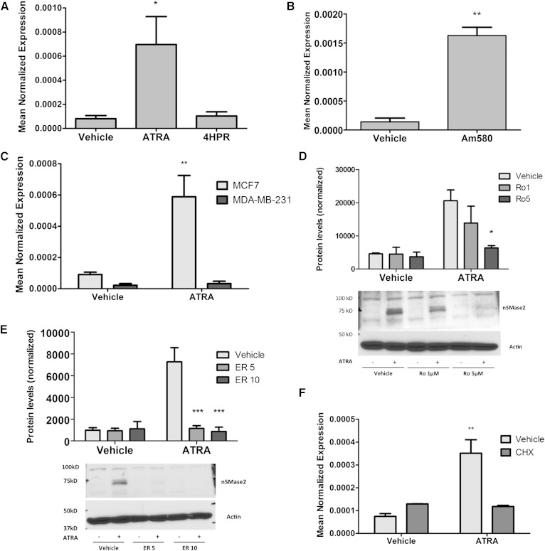
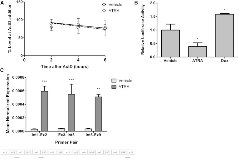
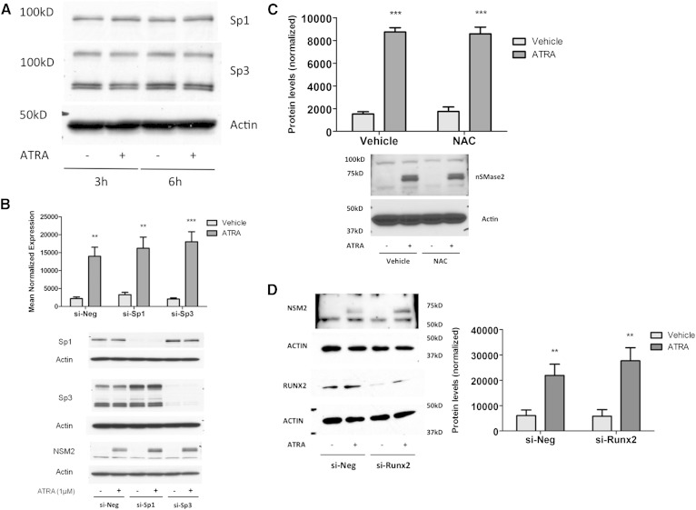
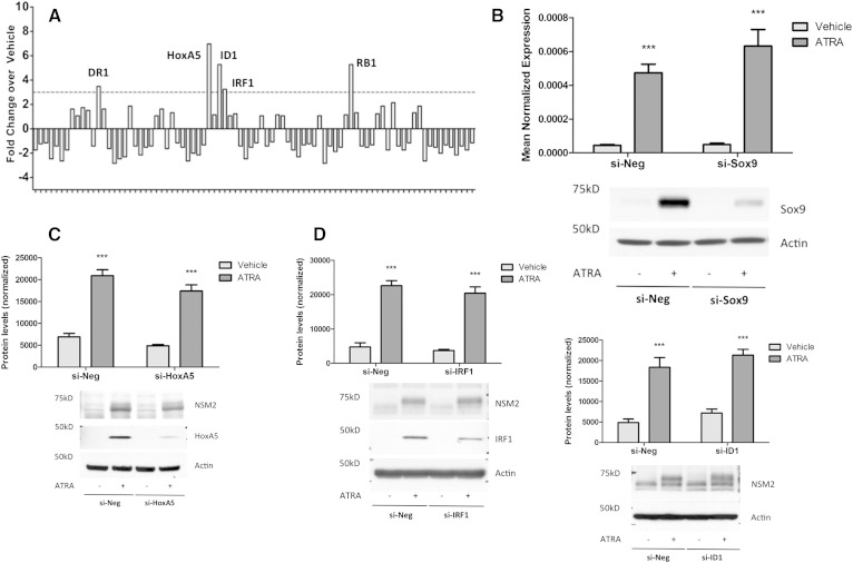
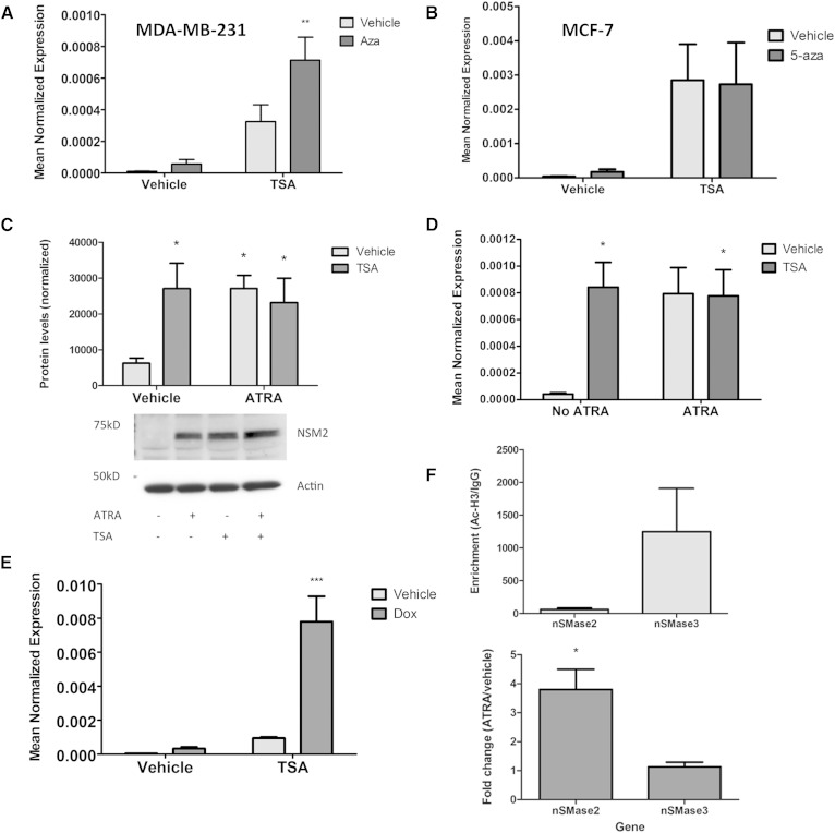
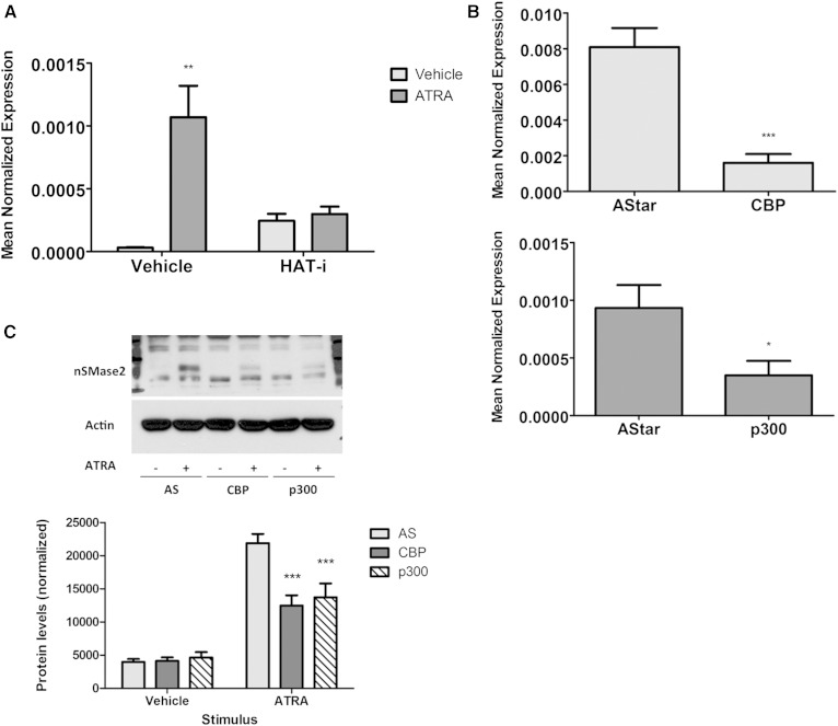
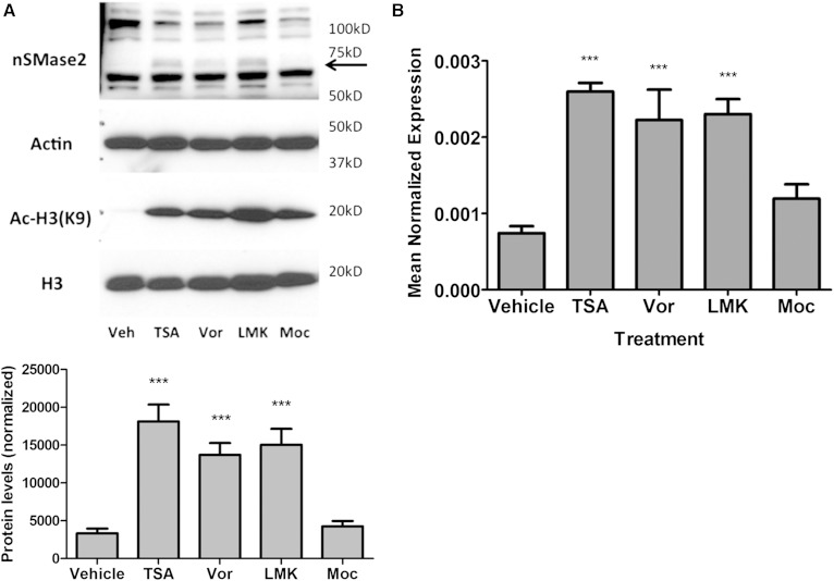
Neutral sphingomyelinase-2 (nSMase2) is a key ceramide-producing enzyme in cellular stress responses. While many posttranslational regulators of nSMase2 are known, emerging evidence suggests a more protracted regulation of nSMase2 at the transcriptional level. Previously, we reported that nSMase2 is induced by all-trans retinoic acid (ATRA) in MCF7 cells and implicated nSMase2 in ATRA-induced growth arrest. Here, we further investigated how ATRA regulates nSMase2. We find that ATRA regulates nSMase2 transcriptionally through the retinoic acid receptor-α, but this is independent of previously identified transcriptional regulators of nSMase2 (Sp1, Sp3, Runx2) and is not through increased promoter activity. Epigenetically, the nSMase2 gene is not repressively methylated in MCF7 cells. However, inhibition of histone deacetylases (HDACs) with trichostatin A (TSA) induced nSMase2 comparably to ATRA; furthermore, combined ATRA and TSA treatment was not additive, suggesting ATRA regulates nSMase2 through direct modulation of histone acetylation. Confirming this, the histone acetyltransferases CREB-binding protein and p300 were required for ATRA induction of nSMase2. Finally, use of class-specific HDAC inhibitors suggested that HDAC4 and/or HDAC5 are negative regulators of nSMase2 expression. Collectively, these results identify a novel pathway of nSMase2 regulation and suggest that physiological or pharmacological modulation of histone acetylation can directly affect nSMase2 levels.

Keywords: all-trans retinoic acid; epigenetic; histone acetyltransferase; histone deacetylase; neutral sphingomyelinase-2; nuclear receptors; retinoids; signal transduction; sphingolipids; transcription.


Figures

Fig. 1.
Fig. 1.
ATRA effects on nSMase2 are through the RAR-α. MCF7 and MDA-MB-231 cells shown were seeded in 60 mm dishes. A: MCF7 cells were stimulated with vehicle (DMSO), ATRA (1 μM), or 4HPR (1 μM) for 12 h (* P < 0.05 vs. vehicle, n = 4). B: MCF7 cells were stimulated with vehicle (DMSO) or Am580 (100 nM) for 12 h (RNA) (** P < 0.01 vs. vehicle, n = 3 RNA). C: MCF7 and MDA-MB-231 cells were stimulated with vehicle (DMSO) or ATRA (1 μM) for 24 h (** P < 0.01 vs. vehicle, n = 3). D: MCF7 cells were pretreated with Ro-415253 (1 or 5 μM) for 20 min prior to stimulation with DMSO or ATRA (1 μM) for 24 h (* P < 0.05 vs. vehicle, n = 4). E: MCF7 cells were pretreated with ER-50891(5 or 10 μM) for 15 min prior to stimulation with DMSO or ATRA (1 μM) for 24 h (*** P < 0.001 vs. vehicle, n = 4). F: MCF7 cells were pretreated with CHX (50 μg/ml) for 15 min prior to stimulation with vehicle (DMSO) or ATRA (1 μM) for 6 h (** P < 0.01 vs. vehicle, n = 3). As shown, RNA was extracted and converted to cDNA, and nSMase2 expression analyzed by qRT-PCR. As shown, protein was extracted in RIPA buffer, and nSMase2 levels analyzed by immunoblot using actin as loading control.
Fig. 2.
Fig. 2.
ATRA induction of nSMase2 is at the transcriptional level. A: MCF7 cells were seeded in 60 mm dishes and stimulated with vehicle (DMSO) or ATRA (1 μM) for 12 h. At this time, transcription was interrupted with actinomycin D (ActD; 1 μg/ml) for 0, 3, 6, and 9 h as shown. At these times, RNA was extracted and nSMase2 expression analyzed by qRT-PCR (P > 0.1; n = 6). B: MCF7 cells were seeded in 60 mm dishes and, 24 h later, were cotransfected with 1 μg nSMase2 promoter-luciferase and 0.5 μg pEF6-LacZ for 6 h prior to stimulation with vehicle (DMSO) or ATRA (1 μM) for 24 h. Cells were lysed, and luciferase and galactosidase activity were determined as described in Materials and Methods (* P < 0.05 vs. vehicle, n ≥ 3). C: MCF7 cells were seeded in 100 mm dishes and stimulated with vehicle (DMSO) or ATRA (1 μM) for 24 h. hnRNA levels were analyzed as described in Materials and Methods (** P < 0.01, *** P < 0.001 vs. vehicle, n = 3).
Fig. 3.
Fig. 3.
Sp1, Sp3, Runx2, and RNS are not required for ATRA effects on nSMase2. A: MCF7 cells were stimulated with vehicle (DMSO) or ATRA (1 μM) as shown. Protein was extracted and analyzed for Sp1 and Sp3 levels by immunoblot using actin as loading control. Immunoblot is representative of n = 3. B: MCF7 cells were seeded in 60 mm dishes and transfected with AStar, Sp1, or Sp3 siRNA (20 nM) for 48 h prior to stimulation with vehicle (DMSO) or ATRA (1 μM) for 24 h. Protein was extracted and analyzed for nSMase2, Sp1, and Sp3 levels by immunoblot using actin as loading control. Immunoblot is representative of n = 4. C: MCF7 cells were pretreated with NAC (500 μM) for 3 h prior to stimulation with vehicle (DMSO) or ATRA (1 μM) for 24 h. Protein was extracted, and nSMase2 levels analyzed by immunoblot (n = 4). D: MCF7 cells were seeded in 60 mm dishes and transfected with AStar or Runx2 siRNA (20 nM) for 48 h prior to stimulation with vehicle (DMSO) or ATRA (1 μM) for 24 h. Protein was extracted and analyzed for nSMase2 and Runx2 levels by immunoblot using actin as loading control. Immunoblot is representative of n = 3.
Fig. 4.
Fig. 4.
Early ATRA-induced transcription factors are not upstream of nSMase2. A: MCF7 cells were seeded in 60 mm dishes and stimulated with vehicle (DMSO) or ATRA (1 μM) for 3 h. RNA was extracted, converted to cDNA, and used as template for the Transcription Factor qRT-PCR array. Data are shown as fold change in ATRA stimulated cells compared with vehicle, n = 1. B: MCF7 cells were transfected with AStar or Sox9 siRNA (20 nM) for 48 h prior to stimulation with vehicle (DMSO) or ATRA (1 μM) for 24 h. Protein was extracted and analyzed for nSMase2 and Sox9 levels by immunoblot using actin as loading control; RNA was extracted and analyzed for nSMase2 by qRT-PCR. Immunoblot is representative of n = 3 (* P < 0.05, n ≥ 3). C: MCF7 cells were transfected with AStar or HoxA5 siRNA (20 nM) for 48 h prior to stimulation with vehicle (DMSO) or ATRA (1 μM) for 24 h. Protein was extracted and analyzed for nSMase2 and HoxA5 levels by immunoblot using actin as loading control. Immunoblot is representative of n = 3. D: MCF7 cells were transfected with AStar or IRF1 siRNA (20 nM) for 48 h prior to stimulation with vehicle (DMSO) or ATRA (1 μM) for 24 h. Protein was extracted and analyzed for nSMase2 and Sox9 levels by immunoblot using actin as loading control. Immunoblot is representative of n = 3. E: MCF7 cells were transfected with AStar or ID1 siRNA (20 nM) for 48 h prior to stimulation with vehicle (DMSO) or ATRA (1 μM) for 24 h. RNA and protein were extracted and analyzed for ID1 (RNA) and nSMase2 (protein) levels by qRT-PCR and immunoblot respectively. Actin was used as reference gene and loading control. Immunoblot is representative of n = 3 (* P < 0.05, n = 3).
Fig. 5.
Fig. 5.
ATRA regulates nSMase2 epigenetically through histone acetylation but not gene methylation. MDA-MB-231 (A) or MCF7 (B) cells were seeded in 60 mm dishes and treated with vehicle or 5-aza (1 μM) for 48 h prior to treatment with vehicle (DMSO) or TSA (300 nM) for 24 h. RNA was extracted and nSMase2 levels analyzed by qRT-PCR using actin as reference gene (** P < 0.01, n = 5). C: MCF7 cells were treated with vehicle (DMSO), ATRA (1 μM), TSA (300 nM), or both for 24 h. Protein lysates were prepared and nSMase2 levels analyzed by immunoblot using actin as loading control (* P < 0.05, n = 3). D: MCF7 cells were treated with vehicle (DMSO), ATRA (1 μM), TSA (300 nM), or both for 24 h. RNA was extracted, and nSMase2 levels were analyzed by qRT-PCR using actin as reference gene (* P < 0.05, n = 3). E: MCF7 cells were treated with vehicle (DMSO), doxorubicin (200 nM), TSA (300 nM), or both for 24 h. RNA was extracted, and nSMase2 levels were analyzed by qRT-PCR using actin as reference gene (** P < 0.01, n = 4). F: MCF7 cells were treated with vehicle (DMSO) or ATRA (1 μM, 24 h), and acetyl-H3 levels at the nSMase2 and nSMase3 genes were probed by ChIP analysis (* P < 0.05, n = 4).
Fig. 6.
Fig. 6.
Knockdown of CBP and p300 blunts ATRA induction of nSMase2. A: MCF7 cells were pretreated with HAT-i (10 μM) for 15 min prior to treatment with vehicle (DMSO) or ATRA (1 μM) for 24 h. RNA was extracted, and nSMase2 levels were analyzed by qRT-PCR using actin as reference gene (* P < 0.05, n = 4). B, C: MCF7 cells were seeded in 60 mm dishes and reverse-transfected with AStar (AS), CBP, or p300 siRNA (20 nM) for 48 h prior to stimulation with vehicle (DMSO) or ATRA (1 μM) for 24 h. B: RNA was extracted with CBP and p300 levels analyzed by qRT-PCR using actin as reference gene (* P < 0.05, *** P < 0.001 vs. AS, n = 4). C: Protein was extracted and analyzed for nSMase2 levels by immunoblot using actin as loading control (*** P < 0.001 vs. AS, n = 4).
Fig. 7.
Fig. 7.
Class II HDACs negatively regulate nSMase2 expression. MCF7 cells were seeded into 60 mm dishes and stimulated with vehicle (DMSO), TSA (300 nM), vorinostat (10 μM), LMK (5 μM), and mocestinostat (5 μM) for 24 h. A: Proteins were extracted in RIPA buffer and analyzed for Ac-H3 (K9), H3, and nSMase2 levels by immunoblot. Actin was used as loading control (*** P < 0.001, n = 4). B: RNA was extracted and converted to cDNA, and nSMase2 expression analyzed by qRT-PCR using actin as reference gene (*** P < 0.001, n = 5).

Similar articles

Cited by

References

    1. Hannun Y. A., and Obeid L. M.. 2008. Principles of bioactive lipid signalling: lessons from sphingolipids. Nat. Rev. Mol. Cell Biol. 9: 139–150. - PubMed
    1. Wu B. X., Clarke C. J., and Hannun Y. A.. 2010. Mammalian neutral sphingomyelinases: regulation and roles in cell signalling responses. Neuromolecular Med. 12: 320–330. - PMC - PubMed
    1. Jenkins R. W., Canals D., and Hannun Y. A.. 2009. Roles and regulation of secretory and lysosomal acid sphingomyelianase. Cell. Signal. 21: 836–846. - PMC - PubMed
    1. Chipuk J. E., McStay G. P., Bharti A., Kuwana T., Schafer B., Clarke C. J., Siskind L. J., Obeid L. M., and Green D. R.. 2012. Sphingolipid metabolism cooperates with BAK and BAX to promote the mitochondrial pathway of apoptosis. Cell. 148: 988–1000. - PMC - PubMed
    1. Clarke C. J., Mediwala K., Jenkins R. W., Sutton C. A., Tholanikunnel B. G., and Hannun Y. A.. 2011. Neutral sphingomyelinase-2 mediates growth arrest by retinoic acid through modulation of ribosomal S6 kinase. J. Biol. Chem. 286: 21565–21576. - PMC - PubMed

Publication types

MeSH terms