. 2016 Apr 1;30(7):856-69.
doi: 10.1101/gad.276428.115. Epub 2016 Mar 24.

Distinct roles of autophagy-dependent and -independent functions of FIP200 revealed by generation and analysis of a mutant knock-in mouse model

Affiliations

Distinct roles of autophagy-dependent and -independent functions of FIP200 revealed by generation and analysis of a mutant knock-in mouse model

Song Chen et al. Genes Dev. .

Abstract

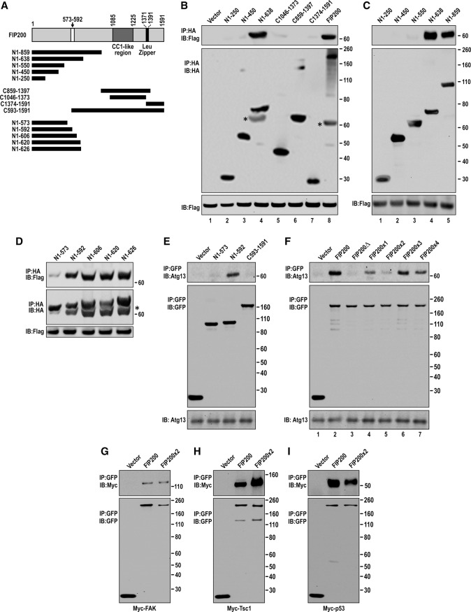
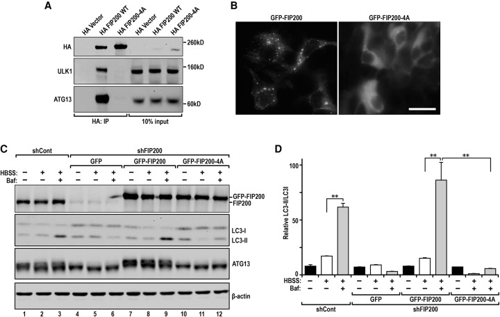
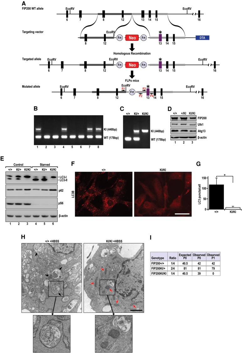
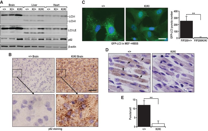
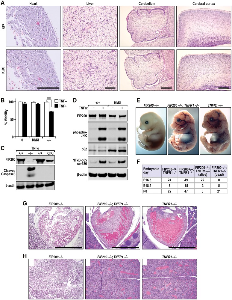
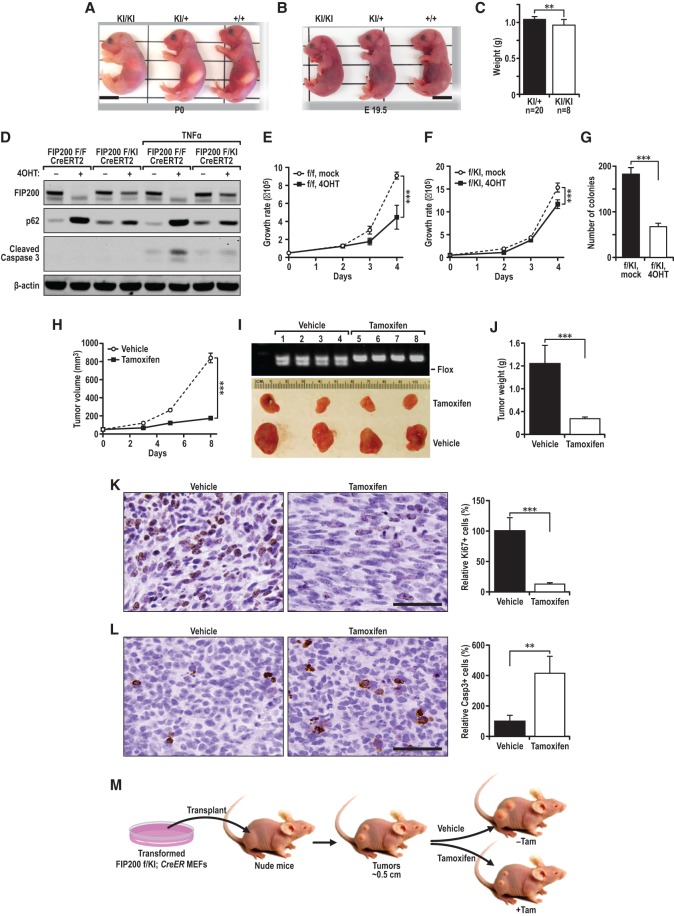
Autophagy is an evolutionarily conserved cellular process controlled through a set of essential autophagy genes (Atgs). However, there is increasing evidence that most, if not all, Atgs also possess functions independent of their requirement in canonical autophagy, making it difficult to distinguish the contributions of autophagy-dependent or -independent functions of a particular Atg to various biological processes. To distinguish these functions for FIP200 (FAK family-interacting protein of 200 kDa), an Atg in autophagy induction, we examined FIP200 interaction with its autophagy partner, Atg13. We found that residues 582-585 (LQFL) in FIP200 are required for interaction with Atg13, and mutation of these residues to AAAA (designated the FIP200-4A mutant) abolished its canonical autophagy function in vitro. Furthermore, we created a FIP200-4A mutant knock-in mouse model and found that specifically blocking FIP200 interaction with Atg13 abolishes autophagy in vivo, providing direct support for the essential role of the ULK1/Atg13/FIP200/Atg101 complex in the process beyond previous studies relying on the complete knockout of individual components. Analysis of the new mouse model showed that nonautophagic functions of FIP200 are sufficient to fully support embryogenesis by maintaining a protective role in TNFα-induced apoptosis. However, FIP200-mediated canonical autophagy is required to support neonatal survival and tumor cell growth. These studies provide the first genetic evidence linking an Atg's autophagy and nonautophagic functions to different biological processes in vivo.

Keywords: FIP200; autophagy; embryogenesis; knock-in mutant mouse; tumor growth.


Figures

Figure 1.
Figure 1.
Identification of residues 582–585 LQFL in FIP200, required for its interaction with Atg13 and autophagy induction. (A) Schematic of the FIP200 fragments used in BE. A white box marks residues 573–592, required for Atg13 binding. (BD) HEK293T cells were cotransfected with expression vectors encoding Flag-tagged Atg13 and HA-tagged FIP200 or its segments or vector alone as a control as indicated. Lysates were prepared and immunoprecipitated by anti-HA agarose beads followed by immunoblotting with antibodies as indicated. (E) The GFP-tagged FIP200 N-terminal or C-terminal segments indicated in A were transfected into HEK293T cells. The lysates were prepared and immunoprecipitated by anti-GFP antibody followed by immunoblotting with antibodies as indicated. (F) HEK293T cells were transfected with expression vectors encoding GFP-tagged-FIP200 or its mutants as indicated. Lysates were prepared and immunoprecipitated by anti-GFP followed by immunoblotting with antibodies as indicated. (GI) GFP-tagged FIP200 or FIP200-4A mutant was cotransfected with Myc-FAK (G), Myc-Tsc1 (H), or Myc-P53 (I), respectively, into HEK293T cells. Lysates were prepared and immunoprecipitated with anti-GFP antibody followed by immunoblotting using antibodies as indicated. Please note that the bands marked by asterisks in the bottom panels of B and D are Flag-tagged Atg13 that was not stripped after immunoblotting using anti-Flag. Molecular weight markers (kilodaltons) are shown at the right.
Figure 2.
Figure 2.
The FIP200-4A mutant unable to bind ATG13 is defective in amino acid starvation-induced autophagy. (A) HEK293T cells were transfected with expression vectors encoding HA-tagged FIP200 and FIP200-4A mutant or vector alone as a control as indicated. Lysates were prepared and immunoprecipitated by anti-HA followed by immunoblotting using anti-HA (top), anti-ULK1 (middle), or anti-Atg13 (bottom). (B) GFP-FIP200 (left) and GFP-FIP200-4A mutant puncta formation in starved HEK293T cells. (C,D) shRNA FIP200 knockdown HEK293T cells reconstituted with shRNA-resistant GFP-FIP200 or the GFP-FIP200-4A mutant were either left untreated or starved (Hank's balanced salt solution [HBSS]) for 2 h and then analyzed by immunoblotting with the indicated antibodies. (C) BafA1 (100 nM), an inhibitor of lysosome degradation, was included for 2 h in samples as indicated. (D) Quantification of LC3I-to-LC3II conversion in FIP200 knockdown HEK293T cells reconstituted with GFP-FIP200 and GFP-FIP200-4A upon starvation as shown in C. (**) P < 0.01.
Figure 3.
Figure 3.
Disruption of FIP200 interaction with Atg13 blocks autophagy in MEFs. (A) Schematic representation of the FIP200-4A mutant knock-in targeting vector and the targeted allele of the FIP200-4A mutant. Solid boxes represent regions of vector homology with the target locus; thick, numbered boxes represent the exons flanking homology regions. Site-directed mutagenesis was used to introduce the LQFL-to-AAAA mutation at the 582–585 amino acid residues. The asterisk denotes the mutated residues on exon 13. (EcoRV) EcoRV sites; (neo) neomycin-resistant gene cassette; (DTA) diphtheria toxin gene. P1 and P2 are a pair of primers for genotyping. P3 and P4 are a pair of primers for sequencing amplified genomic DNA with mutations. (B) Genomic DNA was extracted from mouse tails and analyzed by PCR using P1 and P2 to detect the FIP200 wild-type allele (178 base pairs [bp]) or knock-in allele (448 bp). Representative genotypes of a litter of mice from crosses of FIP200+/KI mice with wild-type B6 mice. (C) Representative genotypes of MEF cells prepared from embryonic day 13.5 (E13.5) embryos of intercrosses of FIP200+/KI mice. (D) Lysates from different MEFs were analyzed by immunoblotting using antibodies against various proteins as indicated. (E) MEFs were incubated in fresh growth medium (control) or HBSS (starvation) in the presence of 100 nM BafA1 for 2 h. Lysates were then prepared and analyzed by immunoblotting using antibodies against proteins as indicated. (F) FIP200+/+ and FIP200KI/KI MEFs were starved in HBSS for 2 h, fixed, and stained for endogenous LC3B. Representative images are shown. Bar, 10 μm. (G) Quantification of LC3B puncta number in starved MEF cells. At least 50 cells per experiment were counted. (**) P < 0.01. (H) Representative transmission electron microscopy images of starved MEF FIP200+/+ and MEF FIP200KI/KI cells. Arrows indicate autophagosomal structures (left panel) and mitochondria (right panel), respectively. Bars, 500 nm. (I) Genotypes of progeny from FIP200+/KI intercrosses.
Figure 4.
Figure 4.
FIP200KI/KI homozygous mice are defective in autophagy. (A) Various FIP200KI/KI neonatal brain, liver, and heart tissue lysates were analyzed by immunoblotting with the indicated antibodies. (B) Immunohistochemistry (IHC) staining of p62 of wild-type and homozygous FIP200 knock-in neonatal brains. (C) GFP-LC3 puncta in FIP200 wild-type and FIP200KI/KI MEFs after starvation with HBSS for 2 h. (D) Neonatal muscle sections from mice expressing GFP-LC3 were stained with anti-GFP antibody. (E) The puncta dot structures were quantified. (**) P < 0.01.
Figure 5.
Figure 5.
Normal morphology of FIP200KI/KI pups at P0 and normal heart and liver development in FIP200−/−;TNFR1−/− embryos at E16.5. (A) Histological sections from the hearts, livers, cerebella, and cerebral cortexes of FIP200+/KI (top panels) and FIP200KI/KI (bottom panels) pups at P0 were analyzed by H/E staining. Bars, 500 µm. (B,C) MEFs from FIP200+/+, FIP200−/−, and FIP200KI/KI (knock-in) embryos were treated for 1 d with or without 50 ng/mL TNFα as indicated. They were analyzed for viability by Trypan blue assay (B) or immunoblotting using antibodies as indicated (C). (D) MEFs from FIP200+/+ (wild-type) and FIP200KI/KI (knock-in) embryos were treated for 10 min with or without 50 ng/mL TNFα as indicated. They were analyzed by immunoblotting using antibodies as indicated. (E) Gross examination of whole-mount FIP200−/−, FIP200−/−;TNFR1−/−, and TNFR1−/− embryos at E16.5. (F) Genotypes of progeny from FIP200+/−,TNFR1−/− intercrosses. (G,H) Histological sections from the hearts (G) and livesr (H) of FIP200−/−, FIP200−/−;TNFR1−/−, and TNFR1−/− embryos at E16.5. Bar, 1 mm.
Figure 6.
Figure 6.
The autophagy function of FIP200 is required to support neonatal survival and tumor cell growth in vivo. (A) Representative morphology of a FIP200KI/KI homozygous neonate compared with its wild-type littermates at P0. (B) Representative neonates of the indicated genotypes delivered by cesarean section at E19.5. (C) The mean body weight of FIP200+/KI and FIP200KI/KI mice. (D) MEF FIP200flox/flox or FIP200flox/KI CreERT2 cells (transformed by E1A/Ras) were treated with 10 nM 4-OHT or control for 72 h followed by addition of 50 ng/mL TNFα for 12 h. Cell lysates were then prepared and analyzed by immunoblotting using various antibodies as indicated. (EG). The cells treated with 4-OHT as in D were used for proliferation assay (E,F) and anchorage-independent growth assay in soft agar (G). (HM).Transformed FIP200f/KI;CreERT2 MEFs were injected subcutaneously into athymic nude mice. (H) Animals (n = 4 each) were treated with vehicle control (−TAM [tamoxifen]; dotted line) or TAM (+TAM; solid line) by intraperitoneal (i.p.) injection, and single tumor growth was measured at the indicated time points. Data points represent means of tumor volume. Tumor burden images (I) and average tumor weight (J) at the final time point are shown. Tumor sections were harvested from the recipient mice at the final time point and analyzed by IHC using anti-Ki67 antibody (K) or anti-cleaved caspase 3 antibody (L). (M) Schematics for the deletion of floxed FIP200 induced by TAM after transplantation of transformed FIP200f/KI;CreERT2 MEFs as described in the Materials and Methods. Bars, 100 μm. (**) P < 0.01; (***) P < 0.001.

References

    1. Abbi S, Guan JL. 2002. Focal adhesion kinase: protein interactions and cellular functions. Histol Histopathol 17: 1163–1171. - PubMed
    1. Boya P, Reggiori F, Codogno P. 2013. Emerging regulation and functions of autophagy. Nat Cell Biol 15: 713–720. - PMC - PubMed
    1. Dillon CP, Weinlich R, Rodriguez DA, Cripps JG, Quarato G, Gurung P, Verbist KC, Brewer TL, Llambi F, Gong YN, et al. 2014. RIPK1 blocks early postnatal lethality mediated by caspase-8 and RIPK3. Cell 157: 1189–1202. - PMC - PubMed
    1. Fan H, Zhao X, Sun S, Luo M, Guan JL. 2013. Function of focal adhesion kinase scaffolding to mediate endophilin A2 phosphorylation promotes epithelial–mesenchymal transition and mammary cancer stem cell activities in vivo. J Biol Chem 288: 3322–3333. - PMC - PubMed
    1. Gan B, Guan JL. 2008. FIP200, a key signaling node to coordinately regulate various cellular processes. Cell Signal 20: 787–794. - PMC - PubMed

Publication types

MeSH terms

Substances