

Effects of short-term exposure to head-down tilt on cerebral hemodynamics: a prospective evaluation of a spaceflight analog using phase-contrast MRI
- PMID: 27013606
- PMCID: PMC4909835
- DOI: 10.1152/japplphysiol.00841.2015
Effects of short-term exposure to head-down tilt on cerebral hemodynamics: a prospective evaluation of a spaceflight analog using phase-contrast MRI
Abstract
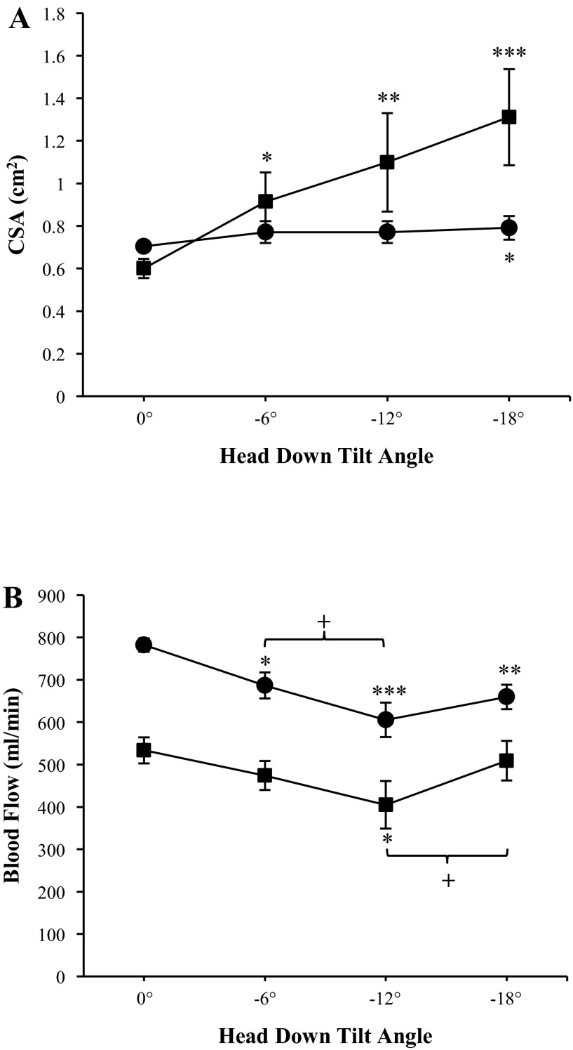
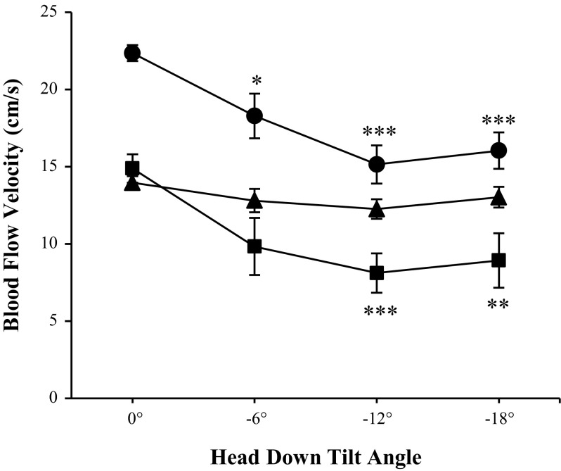
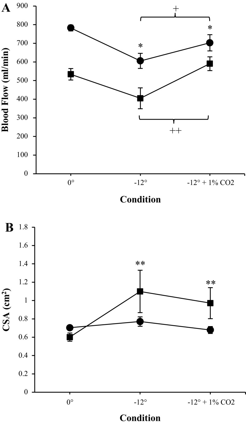
Alterations in cerebral hemodynamics in microgravity are hypothesized to occur during spaceflight and could be linked to the Visual Impairment and Intracranial Pressure syndrome. Head-down tilt (HDT) is frequently used as a ground-based analog to simulate cephalad fluid shifts in microgravity; however, its effects on cerebral hemodynamics have not been well studied with MRI techniques. Here, we evaluate the effects of 1) various HDT angles on cerebral arterial and venous hemodynamics; and 2) exposure to 1% CO2 during an intermediate HDT angle (-12°) as an additional space-related environmental factor. Blood flow, cross-sectional area (CSA), and blood flow velocity were measured with phase-contrast MRI in the internal jugular veins, as well as the vertebral and internal carotid arteries. Nine healthy male subjects were measured at baseline (supine, 0°) and after 4.5 h of HDT at -6°, -12° (with and without 1% CO2), and -18°. We found a decrease in total arterial blood flow from baseline during all angles of HDT. On the venous side, CSA increased with HDT, and outflow decreased during -12° HDT (P = 0.039). Moreover, the addition of 1% CO2 to -12° HDT caused an increase in total arterial blood flow (P = 0.016) and jugular venous outflow (P < 0.001) compared with -12° HDT with ambient atmosphere. Overall, the results indicate decreased cerebral blood flow during HDT, which may have implications for microgravity-induced cerebral hemodynamic changes.
Keywords: MRI; cerebral blood flow; head-down tilt; microgravity; visual impairment and intracranial pressure.
Copyright © 2016 the American Physiological Society.
Figures

References
-
- Alperin N, Lee SH, Sivaramakrishnan A, Hushek SG. Quantifying the effect of posture on intracranial physiology in humans by MRI flow studies. J Magn Reson Imaging 22: 591–596, 2005. - PubMed
-
- Arbeille P, Fomina G, Roumy J, Alferova I, Tobal N, Herault S. Adaptation of the left heart, cerebral and femoral arteries, and jugular and femoral veins during short- and long-term head-down tilt and spaceflights. Eur J Appl Physiol 86: 157–168, 2001. - PubMed
-
- Arbeille P, Provost R, Zuj K, Vincent N. Measurements of jugular, portal, femoral, and calf vein cross-sectional area for the assessment of venous blood redistribution with long duration spaceflight (Vessel Imaging Experiment). Eur J Appl Physiol 115: 2099–2106, 2015. - PubMed
-
- Bagian JP, Hackett P. Cerebral blood flow: comparison of ground-based and spaceflight data and correlation with space adaptation syndrome. J Clin Pharmacol 31: 1036–1040, 1991. - PubMed
-
- Balédent O, Henry-Feugeas MC, Idy-Peretti I. Cerebrospinal fluid dynamics and relation with blood flow: a magnetic resonance study with semiautomated cerebrospinal fluid segmentation. Invest Radiol 36: 368–377, 2001. - PubMed
Publication types
MeSH terms
LinkOut - more resources
Full Text Sources
Other Literature Sources