. 2016 May 20;291(21):11114-23.
doi: 10.1074/jbc.M116.723072. Epub 2016 Mar 24.

The Fragment 1 Region of Prothrombin Facilitates the Favored Binding of Fragment 12 to Zymogen and Enforces Zymogen-like Character in the Proteinase

Affiliations

The Fragment 1 Region of Prothrombin Facilitates the Favored Binding of Fragment 12 to Zymogen and Enforces Zymogen-like Character in the Proteinase

Harlan N Bradford et al. J Biol Chem. .

Abstract

Thrombin is produced from the C-terminal half of prothrombin following its proteolytic activation. The N-terminal half, released as the propiece Fragment 12 (F12), is composed of an N-terminal γ-carboxyglutamate domain (Gla) followed by two kringles (K1 and K2). The propiece plays essential roles in regulating prothrombin activation and proteinase function. The latter results from the ability of F12 to reversibly bind to the (pro)catalytic domain through K2 with high affinity and highly favorable thermodynamic constants when it is a zymogen in comparison to proteinase. Such discrimination is lost for K2 binding after proteolytic removal of the N-terminal Gla-K1 region of F12. The Ca(2+)-stabilized structure of the Gla domain is not required for F12 to bind the zymogen form more favorably. Enhanced binding to zymogen versus proteinase correlates with the ability of the propiece to enforce zymogen-like character in the proteinase. This is evident in variants of meizothrombin, an intermediate of prothrombin activation that contains the propiece covalently attached. This phenomenon is also independent of the Gla domain. Thus, the presence of K1 in covalent linkage with K2 in the propiece governs the ability of K2 to bind the (pro)catalytic domain in favor of zymogen, thereby enforcing zymogen-like character in the proteinase.

Keywords: allosteric regulation; prothrombin; serine protease; thermodynamics; thrombin.


Figures

SCHEME 1.
SCHEME 1.
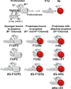
Prothrombin activation and cleaved derivatives. Upper panel, cleavage of prothrombin at Arg320 and Arg271 yields F12 and thrombin (IIa). The propiece, F12, is composed of the N-terminal Gla domain followed by K1 and K2. Thrombin-mediated cleavage at Arg155 separates F12 into the F1 region (Gla-K1) and F2 (K2). The Gla domain can be released by chymotryptic cleavage at Tyr43. The folded structure of the Gla domain is destabilized by EDTA or by the lack of γ-carboxyglutamate modifications (dG). Lower panel, after single cleavage at Arg271, the procatalytic domain (P2) can bind reversibly to the indicated derivatives of the propiece through K2. Thrombin produced by cleavage at both Arg271 and Arg320 can also bind propiece derivatives through K2. The (pro)catalytic domain is shown in gray for the zymogen and in red for the proteinase. Cleavage at Arg320 but not Arg271 yields the proteinase mIIa with the indicated propiece derivatives covalently attached.
SCHEME 2.
SCHEME 2.
Reaction of mIIa-ΔF1 with active site probe (P). The proteinase is proposed to exist in a pre-equilibrium between e and E, of which E binds P with high affinity to form E.P resulting in an increase in fluorescence intensity. Probe binding affinity is given by KE,P = k−2/k+2.
FIGURE 1.
FIGURE 1.
Binding of F12 to zymogen or to proteinase in the presence of EDTA. Integrated heats from heat flow traces were obtained by ITC after sequential injections of 288 μm F12 into 19.5 μm P2A195 (●) or 431 μm F12 into 20.2 μm IIaA195 (○). Measurements were performed in 20 mm Hepes, 0.15 m NaCl, 5 mm EDTA, pH 7.4. The lines are drawn according to the fitted constants listed in Table 1.
FIGURE 2.
FIGURE 2.
Stopped-flow traces of the binding of I-2581 to dG-mIIaQQQ and mIIa-ΔF1. Fluorescence was measured using λEX = 280 nm and λEM >500 nm after rapid mixing of dG-mIIaQQQ (upper trace) or mIIa-ΔF1 (lower trace) with I-2581 in 20 mm Hepes, 0.15 m NaCl, 5 mm CaCl2, 5 μm CoCl2, 0.1% (w/v) PEG8000, pH 7.4, at 25 °C. The final concentrations of proteinase and I-2581 were 0.3 μm and 2 μm. The lower trace is fit to a single exponential rise with Offset = 1.87, Amp = 1.9 ± 0.01, and kobs = 86.5 ± 0.35 s−1. The upper trace is fitted according to a 3 exponential rise with Offset = 1.74, Amp1 = 1.43 ± 0.02, kobs,1 = 96.8 ± 1.8 s−1, Amp2 = 1.11 ± 0.02, kobs,2 = 25.6 ± 0.5 s−1, Amp3 =1.29 ± 0.01, kobs,3 = 1.7 ± 0.01 s−1.
FIGURE 3.
FIGURE 3.
Kinetics of I-2581 binding to mIIa-ΔF1. Main panel, fluorescence traces were obtained after rapid mixing of mIIa-ΔF1 with increasing concentrations of I-2581, and averages of 10–20 replicates are illustrated. Final concentrations were 0.3 μm proteinase and 1, 2, 4, 6, and 8 μm I-2581 (bottom to top). Inset, probe dissociation kinetics was measured by mixing equal volumes of a mixture containing 0.6 μm mIIa-ΔF1 and 4 μm I-2581 with 800 μm FPRck. The black lines in both panels are drawn according to a global fit using Scheme 2 and the fitted constants listed in Table 2. A subset of traces used for global analysis is shown for clarity.
SCHEME 3.
SCHEME 3.
Reaction of other mIIa forms with active site probe (P). The proteinase is proposed to exist in pre-equilibrium between e and E. P binds E with high affinity to form E.P, whereas binding of P to e is of weaker affinity. The fluorescence yields of e.P and E.P are assumed to be identical. Interconversion between e.P and E.P is described by KConf = e.P/E.P. Probe binding affinities are given by KE,P = k−2/k+2 and Ke,P = k−3/k+3.
FIGURE 4.
FIGURE 4.
Kinetics of I-2581 binding to dG-mIIaQQQ. Fluorescence traces are illustrated for the same experimental conditions listed in Fig. 3, except the proteinase was dG-mIIaQQQ. The black lines in both panels are drawn according to a global fit using Scheme 3 and the fitted constants listed in Table 2. A subset of traces used for global analysis is shown for clarity.
FIGURE 5.
FIGURE 5.
Thermodynamics of propiece binding and the zymogen-like character of mIIa. The fractional distribution of mIIa variants in the zymogen-like state inferred from rapid kinetic measurements is illustrated (left axis, black bars). Mean values ± S.E. for fe were taken from Table 2. The values for carboxylated mIIa were taken from Bradford and Krishnaswamy (15). The red bars scaled to the right axis illustrate the difference in enthalpy (ΔΔH) for the interaction of the relevant propiece under appropriate conditions to P2 and to thrombin, taken from Table 1.
FIGURE 6.
FIGURE 6.
Kinetics of I-2581 binding to mIIaQQQ-Δ43. Fluorescence traces are illustrated for the same experimental conditions listed in Fig. 3, except the proteinase was dG-mIIaQQQ-Δ43. The black lines in both panels are drawn according to a global fit using Scheme 3 and the fitted constants listed in Table 2. A subset of traces used for the global analysis is shown for clarity.

References

    1. Mann K. G., Jenny R. J., and Krishnaswamy S. (1988) Cofactor proteins in the assembly and expression of blood clotting enzyme complexes. Annu. Rev. Biochem. 57, 915–956 - PubMed
    1. Mann K. G., Nesheim M. E., Church W. R., Haley P., and Krishnaswamy S. (1990) Surface-dependent reactions of the vitamin K-dependent enzyme complexes. Blood 76, 1–16 - PubMed
    1. Mann K. G. (2003) Thrombin formation. Chest 124, 4S–10S - PubMed
    1. Jenny N. S., Lundblad R. L., and Mann K. G. (2006) Thrombin. In Hemostasis and Thrombosis. Basic Principles and Clinical Practice (Colman R. W., Marder V. J., Clowes A. J., George J. N., and Goldhaber S. Z., eds.) pp 193–213, Lippincott Williams & Wilkins, Philadelphia
    1. Esmon C. T. (2003) The protein C pathway. Chest 124, 26S–32S - PubMed

Publication types

LinkOut - more resources