Comparative Study
. 2016 May 20;291(21):11094-104.
doi: 10.1074/jbc.M115.709139. Epub 2016 Mar 24.

Structure and Metal Binding Properties of a Poxvirus Resolvase

Affiliations
Comparative Study

Structure and Metal Binding Properties of a Poxvirus Resolvase

Huiguang Li et al. J Biol Chem. .

Abstract

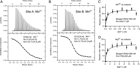
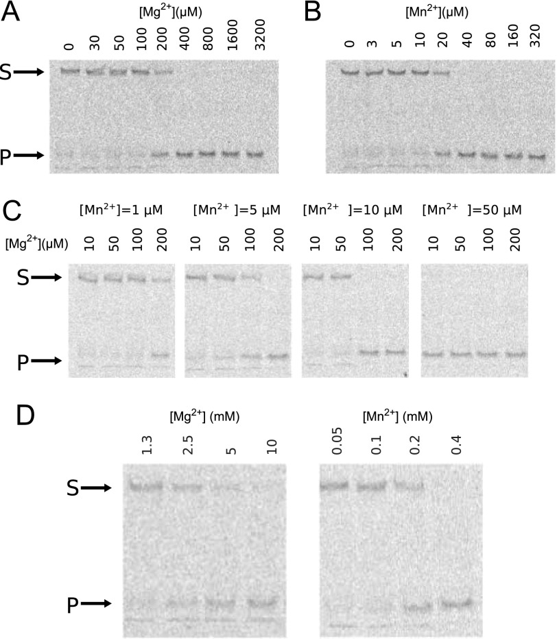
Poxviruses replicate their linear genomes by forming concatemers that must be resolved into monomeric units to produce new virions. A viral resolvase cleaves DNA four-way junctions extruded at the concatemer junctions to produce monomeric genomes. This cleavage reaction is required for viral replication, so the resolvase is an attractive target for small molecule inhibitors. To provide a platform for understanding resolvase mechanism and designing inhibitors, we have determined the crystal structure of the canarypox virus (CPV) resolvase. CPV resolvase is dimer of RNase H superfamily domains related to Escherichia coli RuvC, with an active site lined by highly conserved acidic residues that bind metal ions. There are several intriguing structural differences between resolvase and RuvC, and a model of the CPV resolvase·Holliday junction complex provides insights into the consequences of these differences, including a plausible explanation for the weak sequence specificity exhibited by the poxvirus enzymes. The model also explains why the poxvirus resolvases are more promiscuous than RuvC, cleaving a variety of branched, bulged, and flap-containing substrates. Based on the unique active site structure observed for CPV resolvase, we have carried out a series of experiments to test divalent ion usage and preferences. We find that the two resolvase metal binding sites have different preferences for Mg(2+) versus Mn(2+) Optimal resolvase activity is maintained with 5 μm Mn(2+) and 100 μm Mg(2+), concentrations that are well below those required for either metal alone. Together, our findings provide biochemical insights and structural models that will facilitate studying poxvirus replication and the search for efficient poxvirus inhibitors.

Keywords: Holliday junction; enzyme structure; metalloenzyme; poxvirus; resolvase; viral replication.


Figures

FIGURE 1.
FIGURE 1.
Poxvirus genome replication. Panel i, poxviruses have linear genomes with hairpin ends. ii, rolling hairpin replication results in genome concatemers with inverted repeats (indicated by arrows) located at the concatemer junctions. Panel iii, cruciform extrusion generates a four-way (Holliday) junction at the concatemer junction. Panel iv, the poxvirus resolvase cleaves the junction, resulting in duplex ends that contain single strand breaks (nicks). Panel v, ligation of the nicked duplex ends results in monomeric genome copies.
FIGURE 2.
FIGURE 2.
Overall structure of CPV resolvase. A, orthogonal views of the resolvase dimer. The active sites are indicated by select side chains (yellow/red) and bound Mg2+ (blue spheres). Dimerization is mediated primarily by the α2 helix. Arrows indicate the β4-α2 loop, which adopts different conformations in the two subunits. B, alignment of RuvC family resolvases. Secondary structure is indicated for CPV resolvase. Active site residues are shaded yellow, and residues identical in three or more of the sequences are shaded gray. The α3 helix present in bacterial and mitochondrial resolvases is boxed. Ydc2, Schizosaccharomyces pombe; Tth, T. thermophilus; Ec, E. coli.
FIGURE 3.
FIGURE 3.
A and B, comparison of CPV resolvase (A) and E. coli RuvC (B) structures. The α1 and α2 helices are longer in RuvC, and RuvC has a short α3 helix not present in resolvase. The α4 helix is longer in CPV resolvase. In A, residues whose alanine mutations disrupt activity in FPV resolvase are colored red (Asp8, Glu59, Lys101, Asp131, and Asp134), and those that retain some activity are black (Glu32, Asp54, and Asp130). In B, the conserved active site residues are indicated (yellow/red). C, superposition of resolvase and RuvC subunits. The RuvC structure shown is PDB code 1HJR.
FIGURE 4.
FIGURE 4.
Active site of CPV resolvase. A, stereo view of active site electron density (2mFoDFc; contoured at 1σ) showing the Mg2+-water cluster bound at site A and the five acidic active site residues. B and C, active sites of CPV resolvase (B) and RNase H1 (C) bound to a DNA-RNA hybrid (PDB code 2QKK). Metal coordination and Asp-water hydrogen bonds are drawn as dashed lines. The substrate scissile phosphate is expected to displace one of the water molecules coordinated to Mg2+ in the CPV resolvase A site. Asp130 and Asp134 stabilize a Mg2+-bound water through hydrogen bonding.
FIGURE 5.
FIGURE 5.
Metal ion usage by CPV resolvase. A, direct binding of Mn2+ ions to the CPV resolvase A site by ITC. Mn2+ ions were titrated into a 100 μm solution of CPV resolvase E59A·HJ complex. B, direct binding of Mn2+ ions to the CPV resolvase B site. Mn2+ ions were titrated into a solution of 100 μm CPV resolvase D131A·HJ complex. C, determination of Km for Mn2+ in a CPV resolvase cleavage reaction with bulged DNA substrates. D, determination of Km for Mg2+ in a CPV resolvase cleavage reaction with bulged DNA substrates. In C and D, the concentration of bulged substrate was fixed at the substrate Km values, determined at saturating concentrations of Mn2+ and Mg2+, respectively.
FIGURE 6.
FIGURE 6.
Divalent ion requirements for optimal CPV resolvase activity. A–C, Mg2+ alone as cofactor (A), Mn2+ alone as cofactor (B), and Mn2+ fixed at four different concentrations (C), with Mg2+ varied for each. D, Mg2+ and Mn2+ required for HJ cleavage by the CPV resolvase D130N mutant. The D130N substitution weakens A site metal binding, requiring ∼10-fold higher concentration of either metal ion. The assay in A–D monitors cleavage of 10 nm fluorescently labeled HJ substrate by 100 nm resolvase after 30 min. The arrows indicate the positions of substrate (S) and product (P) bands on native PAGE after quenching the reactions.
FIGURE 7.
FIGURE 7.
Comparison of a CPV resolvase·HJ complex model with the RuvC·HJ complex. A, model of a CPV resolvase·HJ complex obtained by superposition of the resolvase dimer onto the RuvC dimer shown in B. B, structure of the T. thermophilus RuvC·HJ complex determined at 3.8 Å (PDB code 4LD0). The duplex arms of the four-way junction have been extended to 10 bp in length for both A and B. The bound A site Mg2+ ion is drawn as a blue sphere in A, and active site residues are in yellow for both panels. Key regions of protein-DNA interactions discussed in the text are indicated.

References

    1. Moss B. (2001) Poxviridae: the viruses and their replication. In Virology (Fields B. N., ed), pp. 2637–2672, Lippincott-Raven, Philadelphia, PA
    1. Haller S. L., Peng C., McFadden G., and Rothenburg S. (2014) Poxviruses and the evolution of host range and virulence. Infect. Genet. Evol. 21, 15–40 - PMC - PubMed
    1. Moss B. (1991) Vaccinia virus: a tool for research and vaccine development. Science 252, 1662–1667 - PubMed
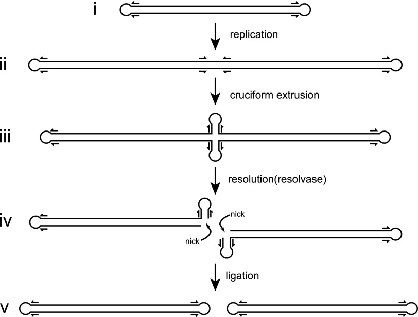
    1. Merchlinsky M., Garon C. F., and Moss B. (1988) Molecular cloning and sequence of the concatemer junction from vaccinia virus replicative DNA. Viral nuclease cleavage sites in cruciform structures. J. Mol. Biol. 199, 399–413 - PubMed
    1. Garcia A. D., Aravind L., Koonin E. V., and Moss B. (2000) Bacterial-type DNA Holliday junction resolvases in eukaryotic viruses. Proc. Natl. Acad. Sci. U.S.A. 97, 8926–8931 - PMC - PubMed

Publication types

MeSH terms

LinkOut - more resources