. 2016 Mar 23;36(12):3453-70.
doi: 10.1523/JNEUROSCI.3693-15.2016.

Dysfunctional Calcium and Glutamate Signaling in Striatal Astrocytes from Huntington's Disease Model Mice

Affiliations

Dysfunctional Calcium and Glutamate Signaling in Striatal Astrocytes from Huntington's Disease Model Mice

Ruotian Jiang et al. J Neurosci. .

Abstract

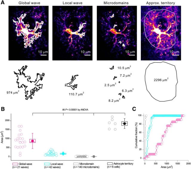
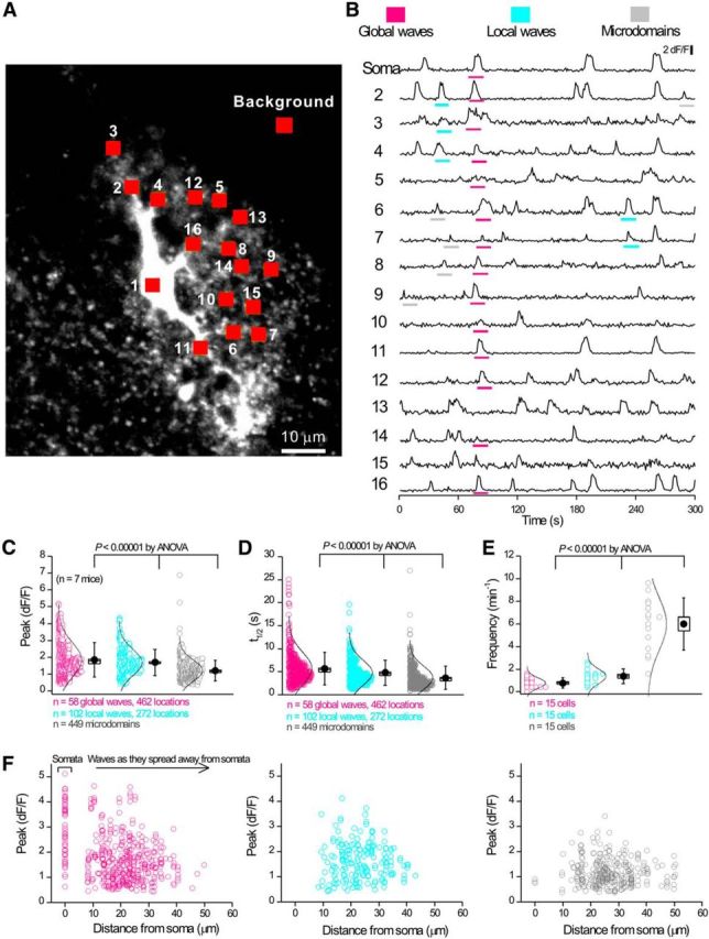
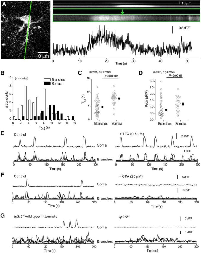
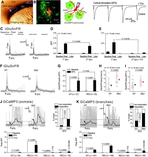
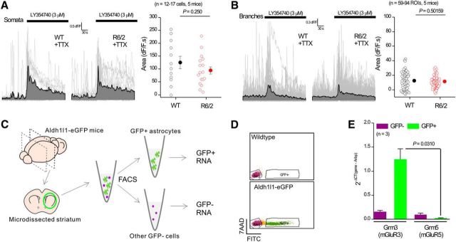
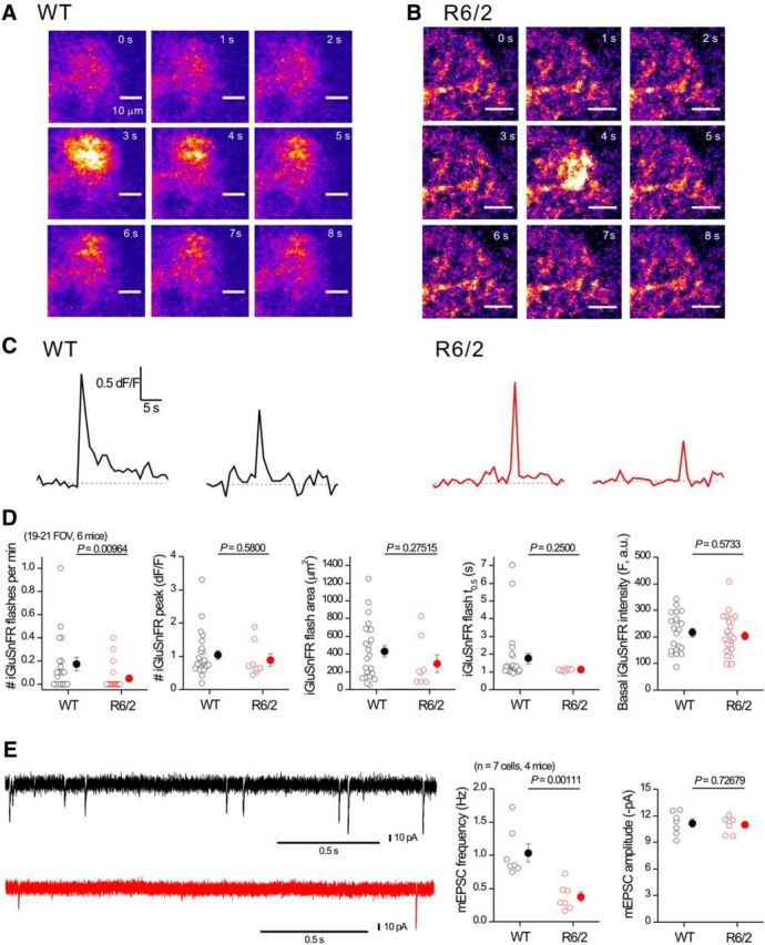
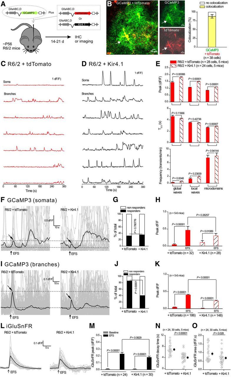
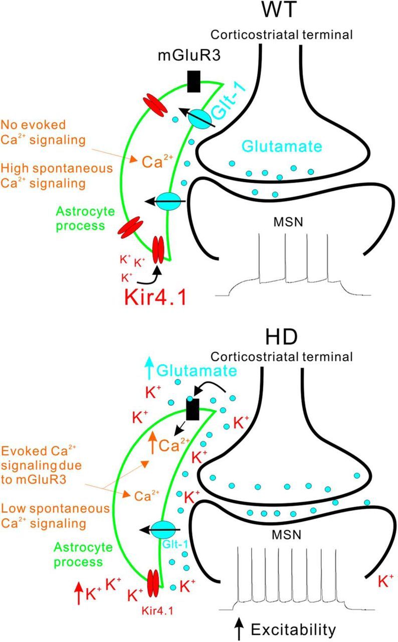
Astrocytes tile the entire CNS, but their functions within neural circuits in health and disease remain incompletely understood. We used genetically encoded Ca(2+)and glutamate indicators to explore the rules for astrocyte engagement in the corticostriatal circuit of adult wild-type (WT) and Huntington's disease (HD) model mice at ages not accompanied by overt astrogliosis (at approximately postnatal days 70-80). WT striatal astrocytes displayed extensive spontaneous Ca(2+)signals, but did not respond to cortical stimulation, implying that astrocytes were largely disengaged from cortical input in healthy tissue. In contrast, in HD model mice, spontaneous Ca(2+)signals were significantly reduced in frequency, duration, and amplitude, but astrocytes responded robustly to cortical stimulation with evoked Ca(2+)signals. These action-potential-dependent astrocyte Ca(2+)signals were mediated by neuronal glutamate release during cortical stimulation, accompanied by prolonged extracellular glutamate levels near astrocytes and tightly gated by Glt1 glutamate transporters. Moreover, dysfunctional Ca(2+)and glutamate signaling that was observed in HD model mice was largely, but not completely, rescued by astrocyte specific restoration of Kir4.1, emphasizing the important contributions of K(+)homeostatic mechanisms that are known to be reduced in HD model mice. Overall, our data show that astrocyte engagement in the corticostriatal circuit is markedly altered in HD. Such prodromal astrocyte dysfunctions may represent novel therapeutic targets in HD and other brain disorders.

Significance statement: We report how early-onset astrocyte dysfunction without detectable astrogliosis drives disease-related processes in a mouse model of Huntington's disease (HD). The cellular mechanisms involve astrocyte homeostasis and signaling mediated by Kir4.1, Glt1, and Ca(2+) The data show that the rules for astrocyte engagement in a neuronal circuit are fundamentally altered in a brain disease caused by a known molecular defect and that fixing early homeostasis dysfunction remedies additional cellular deficits. Overall, our data suggest that key aspects of altered striatal function associated with HD may be triggered, at least in part, by dysfunctional astrocytes, thereby providing details of an emerging striatal microcircuit mechanism in HD. Such prodromal changes in astrocytes may represent novel therapeutic targets.

Keywords: GCaMP; Huntington's disease; astrocyte; calcium.


Figures

Figure 1.
Figure 1.
Three types of striatal astrocyte Ca2+ signals observed in C57BL/6N mice differ in the areas that they cover. A, Representative images and tracings for the areas of global waves, local waves, and microdomains. B, Average data from six mice for experiments and analyses such as those shown in A. Scatter graph shows average area covered by the three types of Ca2+ signals. Statistical comparisons were made using one-way ANOVA. For the box-and-whisker plots in B, the circle represents the mean, the box the SEM, and the whisker the SD. C, Distinct cumulative probability plots for areas covered by global waves, local waves, and microdomains.
Figure 2.
Figure 2.
Striatal astrocytes in C57BL/6N mice display three types of intracellular Ca2+ signals. A, Image of a striatal astrocyte expressing GCaMP3 with 16 ROIs indicated. B, Representative traces from the 16 ROIs shown in A. Global waves, local waves, and microdomains are highlighted in separate colors. CE, Scatter graphs for average data for traces such as those in B show that the three types of Ca2+ signals differ in amplitude, half-width, and frequency. Statistical tests in CE were done using one-way ANOVA. F, Scatter graph of amplitude against distance (from the soma) for the three types of intracellular Ca2+ signals. Local waves and microdomains largely occurred in branches. Statistical comparisons were made using one-way ANOVA. For the box-and-whisker plots in CE, the circle represents the mean, the box the SEM, and the whisker the SD.
Figure 3.
Figure 3.
Properties of intracellular Ca2+ signals in striatal astrocytes in C57BL/6N mice. A, Confocal image of a striatal astrocyte with a green line indicating the approximate position of the region chosen for 200 Hz line scan imaging. Top right, Line scan imaging data for the green line, with an expanded region corresponding to a branch shown below. In these images, the x-axis is time and the y-axis is distance along the scanned line. The trace is plotted for the selected region (framed in green) shown in the recorded image above. B, Distributions showing Ca2+ signal half-widths from line scan experiments for somatic and branch regions. C, D, Scatter plots summarizing Ca2+ signal properties such as amplitude and half-width for line scan data. Statistical tests in C and D were using unpaired Student's t test. E, F, Representative single trace for the soma and three superimposed traces for branches under control conditions and then in the presence of TTX (E) or CPA (F). G, Representative single trace for the soma and three superimposed traces for branches for spontaneous Ca2+ signals recorded from Ip3r2−/− mice and their WT littermates. Average data for TTX, CPA, and Ip3r2−/− experiments (and WT controls) are shown in Table 1. In some cases, the error bars signifying SEM are smaller than the symbols used.
Figure 4.
Figure 4.
Striatal astrocytes in R6/2 mice displayed reduced spontaneous intracellular Ca2+ signals. A, B, Representative traces from eight selected ROIs for spontaneous Ca2+ signals in astrocytes from a WT (A) or from a R6/2 mouse (B). C, Quantification of peak amplitude, duration, and frequency for three types of Ca2+ signals in striatal astrocytes in WT and R6/2 mice (global waves, local waves, and microdomains; see text for further details). D, Schematic showing that CPA blocks SERCA and leads to depletion of intracellular Ca2+ stores due to Ca2+ release from the endoplasmic reticulum (ER). E, Individual traces (gray) and averages (black or red) showing the effect of CPA on astrocyte Ca2+ in WT and R6/2 mice. F, Average data for CPA-evoked Ca2+ increases such as those shown in E. The CPA-mobilized Ca2+ store was smaller in R6/2 mice. In some cases, the error bars signifying SEM are smaller than the symbols used.
Figure 5.
Figure 5.
Striatal astrocytes in R6/2 mice displayed robust Ca2+ signals in response to cortical axon EFS, but those in WT mice did not. A, Photomicrograph of a parasagittal slice of mouse brain showing the position of the stimulating electrode and the imaging site. B, Confocal image (left) and diagram (middle) showing GCaMP3-expressing astrocytes (green) and a MSN filled through the patch-pipette with Alexa Fluor 546. To the right are evoked EPSCs recorded from a MSN in response to 3 EFS; these were abolished in the presence of TTX (0.5 μm). C, Representative traces for a 2-pulse protocol in which 4 EFS were applied twice (first and second stimulation) to cortical axons 10 min apart. Individual traces (gray) and averages (black) for iGluSnFR imaging are shown in response to 4 EFS (arrows). The lines on top of the traces illustrate the time windows in which the peak of the three phases including the baseline, the peak, and later epochs were measured. D, Average data for peak amplitude measured at the three epochs shown in C. E, As in D, with two pulses of 4 EFS at an interval of 10 min, but with the second pulse in the presence of TTX. Note that 2 rounds of 4 EFS produced similar peak responses, whereas interpulse application of TTX abolished the second response. F, Individual traces (gray) and averages (black) for iGluSnFR imaging in response to EFS (arrows) for astrocytes from WT and R6/2 mice. GI, Average data for peak amplitude, area, and kinetics for traces such as those shown in F. J, K, Top left, Individual traces (gray) and averages (black) for GCaMP3 imaging within somata (J) or branches (K) of astrocytes in WT and R6/2 mice, with arrows indicating the time points for EFS and peak response. Note that a delayed slow response was present in both WT and R6/2, but this slow response was not abolished by TTX (see main text). The graphs next to the traces show the percentage of cells responding to EFS. The bar graphs below the traces show average data for peak responses in response to EFS under various conditions for somata and branches. In some cases, the error bars signifying SEM are smaller than the symbols used.
Figure 6.
Figure 6.
Functional and molecular evidence for mGluR3 expression in striatal astrocytes. A, Individual traces (gray) and averages (black) for LY354740-evoked somatic Ca2+ signals within astrocytes for WT and R6/2 mice. The scatter plot to the right shows the average data. B, As in A, but for measurements in branches. C, Schematic for purification of striatal astrocytes from Aldh1l1-eGFP mice at ∼P30 using FACS (see Materials and Methods). D, GFP-negative population (Neg) was defined by the background emission on the FITC channel of GFP-positive cells, and the GFP-positive population was defined by high eGFP fluorescence not overlapping with the GFP-negative cells. E, Bar graph for qPCR analysis of Grm3 and Grm5 from the two purified fractions. The gene expression levels were normalized to the reference gene Arbp using the following formula: 2−ΔCt (Gene of interest-Arbp). In some cases, the error bars signifying SEM are smaller than the symbols used.
Figure 7.
Figure 7.
Blockade of Glt1 in C57BL/6N astrocytes leads to enhanced astrocyte iGluSnFR signals and enhanced astrocyte Ca2+ signals evoked by EFS of cortical axons. A, Individual traces (gray) and averages (black) for GCaMP3 imaging showing increased spontaneous Ca2+ signals in astrocytes from C57BL/6N mice in the presence of TBOA (1 μm). Bar graph to the right shows average data for the peak area before and during applications of TBOA. B, As in A, but for iGluSnFR imaging showing elevated basal glutamate levels in the presence of TBOA. C, Individual traces (gray) and averages (black) for 4 EFS-evoked iGluSnFR signals in the absence of TBOA as well as in its presence. D, E, Average data for peak amplitude and decay time for traces such as those shown in C. F, Individual traces (gray) and averages (black) for 4 EFS-evoked GCaMP3 Ca2+ signals in the presence of TBOA or TBOA/TTX measured in somata of astrocytes in C57BL/6N mice. Tilted arrows indicate the expected time for the EFS-evoked Ca2+ signals. G, Summary bar graph for experiments such as those shown in F in the presence of different drugs. H, I, As in F and G, but for measurements in branches instead of somata. J, Representative confocal images of a GCaMP3-expressing astrocyte taken before, during, and after 4 EFS in the presence of TBOA. Bar graph to the right shows the area of the astrocyte territory and the area of the 4 EFS-evoked Ca2+ signals. In some cases, the error bars signifying SEM are smaller than the symbols used.
Figure 8.
Figure 8.
iGluSnFR flashes are not increased in striatal astrocytes from R6/2 mice. A, Representative time series of confocal images from a small region of an astrocyte expressing iGluSnFR from a WT mouse; an iGluSnFR flash occurred at ∼3 s. B, As in A, but for an astrocyte from a R6/2 mouse. C, Representative traces for transients such as those shown in A and B. D, Bar graphs showing average data for traces such as those shown in C. Note: the only significant difference in iGluSnFR signals between WT and R6/2 mice was in the frequency; no other parameter was changed in R6/2 compared with WT astrocytes. E, Representative traces and average data for mEPSCs recorded from MSNs at −70 mV from WT and R6/2 mice. In some cases, the error bars signifying SEM are smaller than the symbols used.
Figure 9.
Figure 9.
AAV2/5-mediated Kir4.1 expression within astrocytes rescued dysfunctional astrocyte Ca2+ and glutamate signaling in R6/2 mice. A, The diagram illustrates the viral constructs used, and that the mice were microinjected at ∼P50–P56 and studied at P70–P80. B, Confocal imaging for GCaMP3 and tdTomato shows colocalization when two viral constructs were coexpressed in striatal astrocytes. C, D, Representative traces from seven selected ROIs showing enhanced spontaneous Ca2+ signals in astrocytes from R6/2 mice that had received AAV2/5 Kir4.1 (D) compared with R6/2 mice receiving AAV2/5 tdTomato (C). E, Quantification of peak amplitude, duration, and frequency for the three types of Ca2+ signals in striatal astrocytes in R6/2 mice receiving AAV2/5 Kir4.1 or AAV2/5 tdTomato. F, Individual traces (gray) and averages (black) for eight EFS-evoked Ca2+ signals within somata of astrocytes in R6/2 mice receiving AAV2/5 Kir4.1 or AAV2/5 tdTomato. Tilted arrows indicate the peak of the response. G, Bar graph showing the percentage of cells responding to EFS. H, Average data for peak amplitude from traces such as those shown in F. IK, As in FH, but for measurements from branches instead of somata. L, Individual traces (gray) and averages (black) for eight EFS-evoked iGluSnFR signals within astrocyte territories from R6/2 mice receiving AAV2/5 Kir4.1 or AAV2/5 tdTomato. MO, Quantification of peak amplitude, area, and kinetics for traces such as those shown in L for iGluSnFR. In some cases, the error bars signifying SEM are smaller than the symbols used.
Figure 10.
Figure 10.
Schematic summary of the major astrocyte changes observed in R6/2 mice at ∼P70. The top and bottom diagrams summarize key aspects of the changes that occur in astrocytes in HD model mice and how they affect MSNs in the dorsolateral striatum (Tong et al., 2014). Only the key features of our findings are schematized and other differences between WT and R6/2 mice are discussed in the text. In the WT striatum, normal expression levels of Kir4.1 and Glt1 maintain low levels of extracellular K+ and glutamate near synapses. As a result, electrical stimulation of corticostriatal axons does not evoke robust mGluR3-mediated astrocyte Ca2+ signals. However, there are abundant spontaneous Ca2+ signals in striatal astrocytes. In HD model mice, expression levels of Kir4.1 and Glt1 are reduced and this has several observable consequences for astrocytes. Reduced Glt1 levels mean that astrocytes display robust mGluR3-mediated Ca2+ signals during stimulation of corticostriatal axons. This gain of evoked Ca2+ signals is accompanied by a loss of spontaneous Ca2+ signals. The elevated glutamate and K+ levels in the extracellular space increase the excitability of MSNs. Although the observations shown in the diagram were robust and highly significant (see text), at this stage, it is not possible to extend our cellular observations and propose a satisfying circuit based mechanism for how astrocytes cause or contribute to HD symptoms, largely because there is no widely accepted circuit based model for neuronal alterations that accompany HD (Waldvogel et al., 2015). Such circuit-based mechanisms are worthy of further investigation, but will require further detailed evaluations of astrocytes and neurons in WT and HD model mice.

References

    1. Adermark L, Lovinger DM. Electrophysiological properties and gap junction coupling of striatal astrocytes. Neurochem Int. 2008;52:1365–1372. doi: 10.1016/j.neuint.2008.02.006. - DOI - PMC - PubMed
    1. Agulhon C, Fiacco TA, McCarthy KD. Hippocampal short- and long-term plasticity are not modulated by astrocyte Ca2+ signaling. Science. 2010;327:1250–1254. doi: 10.1126/science.1184821. - DOI - PubMed
    1. Agulhon C, Sun MY, Murphy T, Myers T, Lauderdale K, Fiacco TA. Calcium signaling and gliotransmission in normal vs. reactive astrocytes. Front Pharmacol. 2012;3:139. - PMC - PubMed
    1. Barres BA. The mystery and magic of glia: a perspective on their roles in health and disease. Neuron. 2008;60:430–440. doi: 10.1016/j.neuron.2008.10.013. - DOI - PubMed
    1. Behrens PF, Franz P, Woodman B, Lindenberg KS, Landwehrmeyer GB. Impaired glutamate transport and glutamate-glutamine cycling: downstream effects of the Huntington mutation. Brain. 2002;125:1908–1922. doi: 10.1093/brain/awf180. - DOI - PubMed

Publication types