. 2016 Mar 15:10:57.
doi: 10.3389/fncel.2016.00057. eCollection 2016.

KIF5C S176 Phosphorylation Regulates Microtubule Binding and Transport Efficiency in Mammalian Neurons

Affiliations

KIF5C S176 Phosphorylation Regulates Microtubule Binding and Transport Efficiency in Mammalian Neurons

Artur Padzik et al. Front Cell Neurosci. .

Abstract

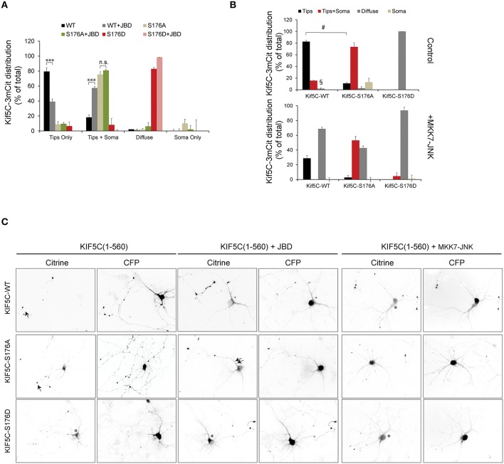
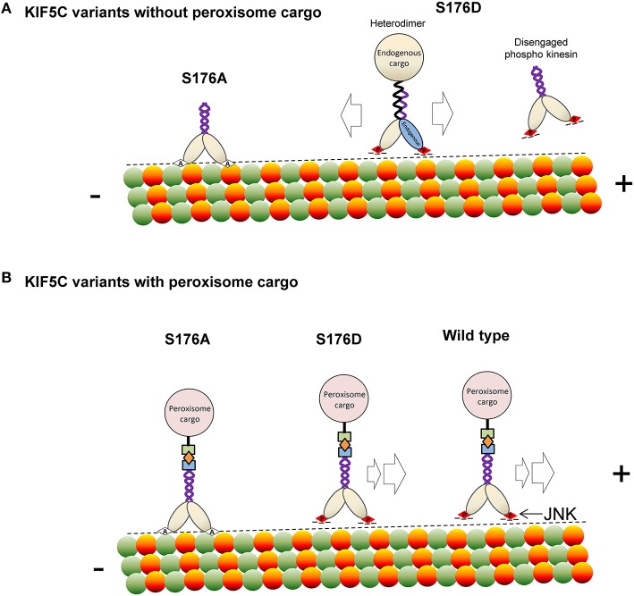
Increased phosphorylation of the KIF5 anterograde motor is associated with impaired axonal transport and neurodegeneration, but paradoxically also with normal transport, though the details are not fully defined. JNK phosphorylates KIF5C on S176 in the motor domain; a site that we show is phosphorylated in brain. Microtubule pelleting assays demonstrate that phosphomimetic KIF5C(1-560)(S176D) associates weakly with microtubules compared to KIF5C(1-560)(WT). Consistent with this, 50% of KIF5C(1-560)(S176D) shows diffuse movement in neurons. However, the remaining 50% remains microtubule bound and displays decreased pausing and increased bidirectional movement. The same directionality switching is observed with KIF5C(1-560)(WT) in the presence of an active JNK chimera, MKK7-JNK. Yet, in cargo trafficking assays where peroxisome cargo is bound, KIF5C(1-560)(S176D)-GFP-FRB transports normally to microtubule plus ends. We also find that JNK increases the ATP hydrolysis of KIF5C in vitro. These data suggest that phosphorylation of KIF5C-S176 primes the motor to either disengage entirely from microtubule tracks as previously observed in response to stress, or to display improved efficiency. The final outcome may depend on cargo load and motor ensembles.

Keywords: BDNF; JNK; SCG10; axonal transport; kinesin; molecular motors; neurons; phosphorylation.


Figures

Figure 1
Figure 1
JNK3 phosphorylates KIF5C on S176, a site that is constitutively phosphorylated in brain. (A) The MS/MS phosphopeptide sequence obtained from in vitro phosphorylation of GST-KIF5C(1-560) by JNK3 shows that S176 (red) contained covalently bound phosphate. MS/MS analysis of phosphopeptides isolated from perinatal mouse brain showed that the same site (S176) was phosphorylated in vivo. Additional KIF5C and KIF5B phosphorylation sites are shown. (B) Kinetics of GST-KIF5C(1-560) phosphorylation by JNK3. Standard errors of the mean (S.E.M.) are shown.
Figure 2
Figure 2
KIF5C(1-560)S176iA moves slower and has a shorter run length than KIF5C(1-560)WT. To determine if S176 affected the speed of KIF5C, single particle tracking of KIF5CWT-3xmCit, KIF5CS176A-3xmCit, KIF5CS176D-3xmCit along taxol-polymerized microtubules was carried out. (A) Time series of KIF5CWT, KIF5CS176A, and KIF5CS176D single particle movements was taken using TIRF microscopy. High resolution arithmetic projections from 300 frames of representative time-lapse movies are shown. Taxol polymerized microtubules are shown in cyan and kinesin particles in red (upper panels) or white (lower panels). The resulting spots do not represent kinesin aggregates, but cumulative presence of a motor at a single coordinate over time. From these projections, it is clear that KIF5CS176A spots are more intense than WT or S176D, indicating longer stationary phases. (B) Next, KIF5C speed was measured from motors moving processively along microtubules. The distribution of speeds for KIF5CWT, KIF5CS176A, and KIF5CS176D are shown. (C) The run length of KIF5C variants is shown. (D) From the same movies, the % of non-motile particles is shown for each of the KIF5C variants. KIF5CS176A shows significantly increased stationary particles. For (A–D), the data was extracted from time-lapse sequences carried out on three separate occasions. The number of particles counted per variant were WT = 207, S176A = 213, and S176D = 86. For (B–D), mean values ± standard error of the means (S.E.M.) from five movies per variant are shown. (E) To visualize the effect of S176 phosphorylation on microtubule binding, COS-7 cells expressing KIF5CWT-3xmCit, KIF5CS176A-3xmCit, or KIF5CS176D-3xmCit were treated with or without AMP-PNP (160 μM) for 10 min. AMP-PNP-treated cells were permeabilized for 1 min using 6 nM streptolysin-O (SLO). Even without addition of AMP-PNP and SLO (left panels), KIFCS176A-3xmCit displayed a filamentous distribution, indicating a prominent association with microtubules (arrows). Scalebar = 10 μm. Significance was determined using Student's t-test. **p < 0.01; ***p < 0.005. (F) Taxol polymerized microtubules were incubated with KIF5C-3xmCit variants as indicated. Microtubules and associated kinesins were pelleted by centrifugation at 100,000 × g, and fractions labeled “S” for supernatant and “P” for pellet, containing microtubule polymers. Fractions were immunoblotted with antibodies detecting KIF5 and tubulin. (G) Quantitative analysis from multiple experiments as described in (F). This data shows that KIF5CS176A-3xmCit associated with the polymerized microtubule fraction (P), even in the absence of AMP-PNP. (H) The pelleting experiments were carried out in the absence of microtubules to determine whether the KIF5CS176A variant pelleted due to aggregate formation. However, in the absence of microtubules, KIF5CS176A pelleting was equivalent to WT and S176D. Meaned data ± S.D. from two separate experiments are shown.
Figure 3
Figure 3
JNK activity regulates the trafficking of KIF5C in hippocampal neurons. (A) Hippocampal neurons at 4 days in vitro were transfected with KIF5C-3xmCit variants, as indicated and CFP (to reveal the cytosolic space). Distribution to neurite tips (tips only), soma and neurites (tips and soma), and neurites (diffuse) was measured. The influence of the JNK inhibitor JBD was also tested. Mean values ± S.E.M. from four independent experiments are shown. (B) Hippocampal neurons at 4 days in vitro were transfected with KIF5C variants as shown, in the presence or absence of the active JNK chimera, MKK7-JNK. (C) Representative images of citrine-tagged (KIF5C variants) and co-expressed CFP are shown. Arrow heads highlight neurite tips with enriched motors and asterisks mark diffuse motor distribution at the soma and in neurites. Mean data ± S.E.M. is shown. ***p < 0.005; §, p < 0.0001; #p < 0.0001 between groups KIF5CWT and KIF5CWT +MKK7-JNK.
Figure 4
Figure 4
Active JNK displaces 50% of KIF5C from microtubules while the remaining 50% displays improved motility and bidirectional movement. (A) The parameters used to filter movements of KIF5C variants in 4–5 day hippocampal neurons are shown schematically. (B) Extrapolated motor run speeds from particle movements are plotted. (C) The percentage of motors exhibiting at least one non-motile event (pausing) during the 10 min acquisition is depicted. (D) Percentage of non-motile motors is shown. This refers to particles that fulfilled the size criteria but demonstrated <1 μm displacement during the entire recording. (E) The percentage of particles showing very shorts runs or diffuse movements is shown. This was defined as those particles recognized within the filter parameters for size and intensity, but lacking the minimum five connected frames (A). Approximately 50% of particles in neurons expressing either KIF5CS176D or KIF5CWT in the presence of MKK7-JNK, displayed these characteristics. (F) Kymographs of KIF5C(1-560) variant movements over 10 min. (G) Maximum intensity projections of straightened axons from hippocampal neurons transfected with the KIF5C variants. (H) Directionality of motor movement, retrograde, anterograde, or bidirectional, is shown (mean data ± S.E.M.). The number of experiments (n) is indicated and the number of tracked particles is shown in parenthesis for each KIF5 variant: KIF5CWT n = 7, (389); KIF5CS176A, n = 5 (307); KIF5CS176D, n = 6 (139); KIF5CWT +JBD, n = 7 (235); KIF5CWT +MKK7-JNK, n = 7 (312). (I) The effect of JNK on KIF5(1-376)WT-catalyzed ATP hydrolysis was measured in the presence of polymerized microtubules following 30 min pre-incubation with active JNK. Kinase activity was inhibited during the ATP hydrolysis reaction using 1 μM SP600125. JNK phosphorylation increased ATP hydrolysis by KIF5CWT. (J) KIF5C ATPase activity was measured in the absence of microtubules. Averaged data from three experiments is shown. Standard errors of the mean are shown for each data point. Statistical analysis was by two-way ANOVA and Bonferroni post hoc test. Error bars represent S.E.M.s. Significance levels, *p < 0.05; **p < 0.005; ***p < 0.0005; #p < 0.0001.
Figure 5
Figure 5
KIF5CS176A-mediated cargo transport is reduced in living cells. COS-7 cells were transfected with PEX-mRFP-FKBP and KIF5CWT-GFP-FRB or KIF5CS176A-GFP-FRB or KIF5CS176D-GFP-FRB. Time is shown in minutes. (A) Scheme of the inducible cargo trafficking assay. Rapalog was added at t = 00.00 to induce binding of KIF5C constructs to peroxisomes. (B) Localization of KIF5C mutants in COS-7 cells co-expressing PEX-mRFP-FKBP. Scale bar = 10 μm. (C) Distribution of peroxisomes before (t = -05:00) and after rapalog addition (t = 25.00). Color plots show the spatial displacement of peroxisomes colored according to time with blue frames showing peroxisome localization before rapalog, and red frames after rapalog addition. (D) Kymographs of cells shown in (C). Kymographs were drawn from the cell center to the periphery as indicated in (C). Horizontal and vertical scale bars indicate 10 min and 5 μm respectively. (E) Displacement graphs showing 90% of peroxisome intensity relative to the cell center over time.
Figure 6
Figure 6
KIF5 transport in neurons is regulated by JNK. (A) To test whether SCG10 was a KIF5 cargo, hippocampal neurons were transfected with dominant negative inhibitors of KIF5 transport HA-KLC-TPR or HA-KIF5C(672-955). Endogenous SCG10 or HA-tagged dominant negative inhibitors were detected with α-SCG10 or α-HA antibodies respectively. Inverted micrographs of fluorescence composites are shown. Scalebar = 40 μm. Arrow indicates neurons that were positive for HA. (B) SCG10 distribution in neurons was scored at the cell soma and in growth cones. In neurons expressing dominant inhibitors of KIF5 transport, SCG10 displayed a diffuse signal in the cytosol and no enrichment at the Golgi or in growth cones (*). Mean values ± S.E.M. are shown from four individual experiments. Arrows point to SCG10 immunoreactivity. (C) Micrographs of postnatal day 7 rat brain showing enrichment of SCG10 in nerve tracts (brown). Hematoxylin was used as a counter stain (blue). (D) Transport of Venus-SCG10 and Venus-BDNF in cortical neurons in the presence or absence of the JNK inhibitor JBD was monitored using CCD imaging. The speed of vesicles carrying Venus-SCG10 or Venus-BDNF was averaged from multiple time-lapse movies. (E) Distribution plots of Venus-SCG10 transport taken from (D). (F) Distribution plots of Venus-BDNF transport taken from (D). (G) Kymograph plots of movements over 2 min are shown. (H) The % of pausing of Venus-SCG10 or Venus-BDNF is shown. Measured cargo speeds (D–F) do not take into account stationary vesicles. Expression of the JNK inhibitor JBD substantially increased pause frequency of both cargos. (I) The % of non-motile Venus-SCG10 and Venus-BDNF cargos are shown. Averaged data ± S.E.M. is shown. *p < 0.05; **p < 0.01; #p < 0.0001.
Scheme 1
Scheme 1
A model depicting how phosphorylation of KIF5C on S176 may regulate microtubule binding and motility. (A) In the absence of rapalog-tethered cargo, S176 phosphorylation causes either complete disengagement of KIF5C(1-560) from microtubules or increased bidirectional shuffling. In contrast, non-phosphorylated KIF5C(1-560) associates tightly with microtubules. (B) In the peroxisome cargo-bound state, S176 phosphorylated KIF5C(1-560) transports to microtubule plus ends, whereas dephosphorylated KIF5C(1-560) is bound tightly to microtubules resulting in an immobile state. As a consequence, phosphorylation of S176 can facilitate plus-end cargo transport by KIF5C(1-560).

References

    1. Amos L. A., Hirose K. (2007). A cool look at the structural changes in kinesin motor domains. J. Cell Sci. 120, 3919–3927. 10.1242/jcs.016931 - DOI - PMC - PubMed
    1. Arpağ G., Shastry S., Hancock W. O., Tüzel E. (2014). Transport by populations of fast and slow kinesins uncovers novel family-dependent motor characteristics important for in vivo function. Biophys. J. 107, 1896–1904. 10.1016/j.bpj.2014.09.009 - DOI - PMC - PubMed
    1. Baas P. W., Lin S. (2011). Hooks and comets: the story of microtubule polarity orientation in the neuron. Dev. Neurobiol. 71, 403–418. 10.1002/dneu.20818 - DOI - PMC - PubMed
    1. Björkblom B., Ostman N., Hongisto V., Komarovski V., Filén J. J., Nyman T. A., et al. . (2005). Constitutively active cytoplasmic c-Jun N-terminal kinase 1 is a dominant regulator of dendritic architecture: role of microtubule-associated protein 2 as an effector. J. Neurosci. 25, 6350–6361. 10.1523/JNEUROSCI.1517-05.2005 - DOI - PMC - PubMed
    1. Björkblom B., Padzik A., Mohammad H., Westerlund N., Komulainen E., Hollos P., et al. . (2012). c-Jun N-terminal kinase phosphorylation of MARCKSL1 determines actin stability. Mol. Cell. Biol. 32, 3513–3526. 10.1128/MCB.00713-12 - DOI - PMC - PubMed