

Differences in F-Wave Characteristics between Spinobulbar Muscular Atrophy and Amyotrophic Lateral Sclerosis
- PMID: 27014057
- PMCID: PMC4783393
- DOI: 10.3389/fnagi.2016.00050
Differences in F-Wave Characteristics between Spinobulbar Muscular Atrophy and Amyotrophic Lateral Sclerosis
Abstract
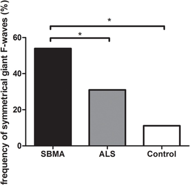
There is limited data on the differences in F-wave characteristics between spinobulbar muscular atrophy (SBMA) and lower motor neuron dominant (LMND) amyotrophic lateral sclerosis (ALS). We compared the parameters of F-waves recorded bilaterally from the median, ulnar, tibial, and deep peroneal nerves in 32 SBMA patients, 37 patients with LMND ALS, and 30 normal controls. The maximum F-wave amplitudes, frequencies of giant F-waves, and frequencies of patients with giant F-waves in all nerves examined were significantly higher in the SBMA patients than in the ALS patients and the normal controls. The mean F-wave amplitude, maximum F-wave amplitude, frequency of giant F-waves, and frequency of patients with giant F-waves in the median and deep peroneal nerves were comparable between the ALS patients and normal controls. Giant F-waves were detected in multiple nerves and were often symmetrical in the SBMA patients compared with the ALS patients. The number of nerves with giant F-waves seems to be the most robust variable for differentiation of SBMA from ALS, with an area under the curve of 0.908 (95% CI: 0.835-0.982). A cut-off value of the number of nerves with giant F-waves (≥3) for diagnosing SBMA showed high sensitivity and specificity: 85% sensitivity and 81% specificity vs. ALS patients. No significant correlations were found between the pooled frequency of giant F-waves and disease duration in the SBMA (r = 0.162, P = 0.418) or ALS groups (r = 0.107, P = 0.529). Our findings suggested that F-waves might be used to discriminate SBMA from ALS, even at early stages of disease.
Keywords: F-wave; amyotrophic lateral sclerosis; giant F-wave; motor neuron; nerve conduction study; spinobulbar muscular atrophy.
Figures

Similar articles
-
A Retrospective Study of the Characteristics and Clinical Significance of A-Waves in Amyotrophic Lateral Sclerosis.Front Neurol. 2017 Sep 28;8:515. doi: 10.3389/fneur.2017.00515. eCollection 2017. Front Neurol. 2017. PMID: 29033889 Free PMC article.
-
[Sample size for the estimation of F-wave parameters in healthy volunteers and amyotrophic lateral sclerosis patients].Zhonghua Yi Xue Za Zhi. 2017 Mar 7;97(9):670-674. doi: 10.3760/cma.j.issn.0376-2491.2017.09.007. Zhonghua Yi Xue Za Zhi. 2017. PMID: 28297826 Chinese.
-
Discrimination of spinal and bulbar muscular atrophy from amyotrophic lateral sclerosis using sensory nerve action potentials.Muscle Nerve. 2012 Feb;45(2):169-74. doi: 10.1002/mus.22291. Muscle Nerve. 2012. PMID: 22246870
-
The genetics of motor neuron diseases.Amyotroph Lateral Scler Other Motor Neuron Disord. 2003 Dec;4(4):225-31. doi: 10.1080/14660820310011287. Amyotroph Lateral Scler Other Motor Neuron Disord. 2003. PMID: 14753656 Review.
-
Motor Neuron Diseases and Neuroprotective Peptides: A Closer Look to Neurons.Front Aging Neurosci. 2021 Sep 17;13:723871. doi: 10.3389/fnagi.2021.723871. eCollection 2021. Front Aging Neurosci. 2021. PMID: 34603008 Free PMC article. Review.
Cited by
-
Unique clinical and electrophysiological features in the peripheral nerve system in patients with sialidosis - a case series study.Orphanet J Rare Dis. 2024 May 24;19(1):217. doi: 10.1186/s13023-024-03216-8. Orphanet J Rare Dis. 2024. PMID: 38790028 Free PMC article.
-
F-Wave Features in Most Common Chinese Spinocerebellar Ataxias.Cerebellum. 2024 Dec 10;24(1):8. doi: 10.1007/s12311-024-01753-3. Cerebellum. 2024. PMID: 39658703
-
A Retrospective Study of the Characteristics and Clinical Significance of A-Waves in Amyotrophic Lateral Sclerosis.Front Neurol. 2017 Sep 28;8:515. doi: 10.3389/fneur.2017.00515. eCollection 2017. Front Neurol. 2017. PMID: 29033889 Free PMC article.
-
Clinical and Physiological Significance of F-Wave in Spinocerebellar Ataxia Type 3.Front Neurol. 2020 Sep 29;11:571341. doi: 10.3389/fneur.2020.571341. eCollection 2020. Front Neurol. 2020. PMID: 33117264 Free PMC article.
-
The French national protocol for Kennedy's disease (SBMA): consensus diagnostic and management recommendations.Orphanet J Rare Dis. 2020 Apr 10;15(1):90. doi: 10.1186/s13023-020-01366-z. Orphanet J Rare Dis. 2020. PMID: 32276665 Free PMC article.
References
-
- Baumann F., Henderson R. D., Ridall P. G., Pettitt A. N., McCombe P. A. (2012). Quantitative studies of lower motor neuron degeneration in amyotrophic lateral sclerosis: evidence for exponential decay of motor unit numbers and greatest rate of loss at the site of onset. Clin. Neurophysiol. 123 2092–2098. 10.1016/j.clinph.2012.03.007 - DOI - PubMed
LinkOut - more resources
Full Text Sources
Other Literature Sources
Miscellaneous