Review
. 2016 Mar 10:7:275.
doi: 10.3389/fpsyg.2016.00275. eCollection 2016.

Links of Consciousness, Perception, and Memory by Means of Delta Oscillations of Brain

Affiliations
Review

Links of Consciousness, Perception, and Memory by Means of Delta Oscillations of Brain

Erol Başar et al. Front Psychol. .

Abstract

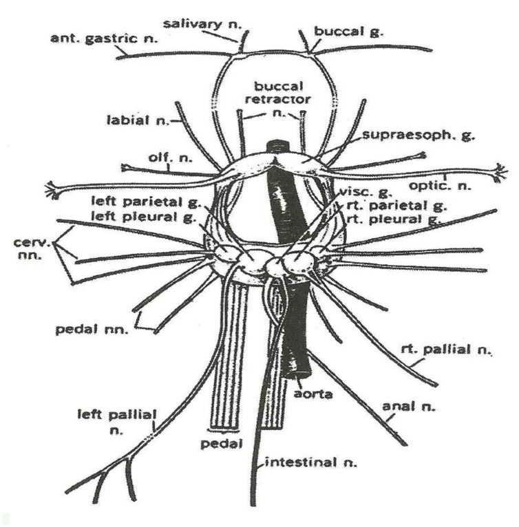
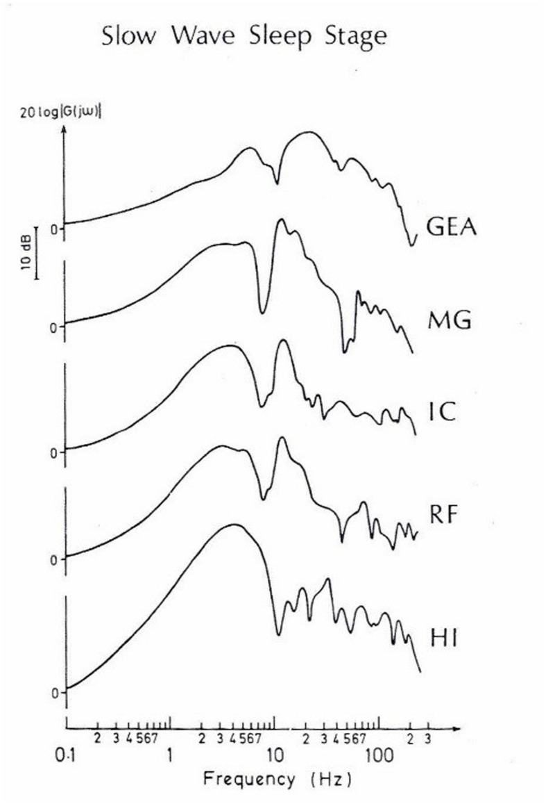
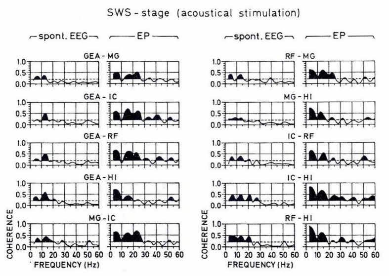
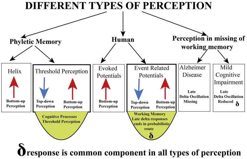
The aim of this report is threefold: (1) First, we accomplish a survey integrating the description of consciousness, perception, and memory according to the views of descriptions of Hermann Helmholtz, Sigmund Freud, Henri Bergson, and Gustav Jung. (2) In the second step, we present experimental results for defining the machineries of sensation and perception: (a) electrical responses of isolated ganglion of Helix pomatia were measured upon odor stimuli that elicited varied degrees of responses. Such a model may give an idea of the control of sensation in the preconscious state of a living tissue. (b) We also describe experiments at the human hearing threshold level. (c) Further, the omission of working memory will be shown with the attenuation of delta response in Alzheimer's subjects in P300 measurements. (d) Finally, the measurement of auditory evoked potentials during slow-wave sleep in the cat brain explains the auditory responses that are not heard at this level of consciousness. (3) In the third step, we aim to provide a synopsis related to integration of perception, memory, and consciousness. By using concepts of important scientists as S. Freud on consciousness, we also tentatively discuss the boundaries of the transition of unconsciousness states to conscious states.

Keywords: P300; brain oscillations; consciousness; perception; top–down; unconsciousness; working memory.


Figures

FIGURE 1
FIGURE 1
In this illustration, past memory includes semantic memory and episodic memory. Semantic memory and episodic memory are mostly overlapping in time, and possibly, they share similar neural networks. Emotional memory is also based on our past memory. Therefore, it is designed in an adjoining way to episodic memory. The illustration also indicates that present memory relies on past memory, and that there possibly are links from past memory to future memory and creativity. As stated by Bergson (1920), the consideration of future events requires past and present memories. As stated in the text, the time space, which we call hypertime space, requires a physical time period of approximately 0.5 s. In this illustration, the functioning of working memory and implicit memory are not yet incorporated. Modified and extended from Başar and Düzgün (2015).
FIGURE 2
FIGURE 2
Explanation see in the text.
FIGURE 3
FIGURE 3
The pink area in this schematic description of neural pathway is involved with bottom–up processing. The gray areas in this illustration are tentative neural pathways, in which complex top–down processing may take place. As indicated with arrows during top–down processing, there are several possible loops generating recurrent reverberations. According to Fessard (1961), it is impossible to predict exactly in which areas the signal transmission ends. The exact duration of transmission is not possible, and this case is dependent on cognitive states. It is also possible to express that the results are probabilistic and also include the unconsciousness inference of Helmholtz.
FIGURE 4
FIGURE 4
Grand average ERPs (50 checkerboards; N = 9 subjects), filtered 1–5 Hz, respectively. (Left) Visual evoked potentials (VEP), (middle) responses to non-target stimuli; (right) responses to target stimuli.
FIGURE 5
FIGURE 5
Mild cognitive impairment (MCI) and Alzheimer’s disease (AD) continuity is prominent in auditory event-related delta oscillatory activity, showing gradually decreasing delta amplitudes and delayed delta peak responses among healthy subjects, MCI, and mild Alzheimer subjects (Modified from Yener et al., 2011).
FIGURE 6
FIGURE 6
The central nervous system and major nerves of a young Helix aspersa (Modified from Courtesy G. Kerkut).
FIGURE 7
FIGURE 7
The power spectra of recordings from the corresponding experiment. Each panel consists of 20 single epochs (204.8 s). From bottom: control (prestimulus), immediately after application of ethanol and the following recordings at different times. The stimulus was removed at 7 min after stimulus onset. Note that fluctuation with multiple frequency components reaching well into 10 Hz and beyond was induced shortly after ethanol, most strongly in the 0.5–5 Hz range (panel 2). Then it subsided to occasional small bursts (panels 3 and 4). Firing continued intermittently even after removal of stimulus (panels 5–7).
FIGURE 8
FIGURE 8
Relationship between degree of aversion and frequency of odorant-induced activity of the Helix pedal ganglion. The bars represent 95% confidence limits for the means. Note that there was a linear relationship between degree of averseness and peak power frequency of odorant-induced FP activity in the Helix ganglion. This diagram suggests: (a) order of averseness appears to reflect that of molecular affinity: the stronger in affinity an odorant, the more aversive it is to the snail; (b) order of affinity is correlated to peak power frequency of induced FP: the stronger in affinity an odorant, the lower the odorant-induced frequency; (c) the more aversive an odorant, the lower the peak power frequency; (d) extrapolation of the curve to the abcissa yields a value of ∼2.5 Hz. This is the area for an appetitive odorant (ethanol) for the snail. These curves suggest that the odorant-specific low frequencies may, together with other frequency components, be involved in identification, classification, and discrimination of odorants or their classes and that the most crucial FP activities relevant for this function may exist below ∼2.5 Hz (Modified from Schütt et al., 1999).
FIGURE 9
FIGURE 9
Grand average auditory evoked potentials recordings of ten subjects (vertex). (A) 0.5–3 Hz digitally filtered AEPs to different stimulation intensities. TH (tone heard) and TNH (tone not heard) are subgroups of the threshold experiments. SPO are the spontaneous activity experiments. (B) The same potentials are reproduced unfiltered. Negativity is upward.
FIGURE 10
FIGURE 10
Grand average peak-to-peak AEP amplitudes of ten subjects (vertex recording). Below are the absolute values, together with standard deviations, above a histogram representation. TH; TNH: both subgroups of threshold stimulation. SPO = spontaneous activity experiments. The evoked potentials to the different stimulation intensities have been digitally filtered in different frequency ranges. The resulting peak-to-peak amplitudes of the digitally filtered activities in different frequency ranges. The resulting peak-to-peak amplitudes of the digitally filtered activities are presented.
FIGURE 11
FIGURE 11
A typical set of amplitude-frequency characteristics obtained by means of the frequency response analyze method and using the selectively averaged evoked potentials (SAEPs), which were simultaneously recorded from different brain nuclei of the cat during the slow-wave sleep stage. Direct computer plottings. Along the abscissa is the input frequency in logarithmic scale, along the ordinate is the potential amplitude, | G(jω)|, in decibels. The curves are normalized in such a way that the amplitude at 0 Hz is equal to 1 (or 20 log 1 = 0). (Modified from Başar et al., 1979).
FIGURE 12
FIGURE 12
A typical set of coherence functions computed from the spontaneous and evoked potentials of all possible pairings of the studied brain structures during the slow-wave sleep stage. The scale is indicated at the bottom. Along the abscissa is the frequency from 0 to 60 Hz, along the ordinate is the coherency between 0 and 1. The horizontal broken lines indicate the significance level, which is 0.2 for all plots. The area under the coherence functions is darkened only if the curve surpasses this level. In order to facilitate a comparison between the coherence values computed from spontaneous and evoked parts of the EEG, the respective coherence functions are presented adjacently as couples for all the pairings of recording electrodes (From Başar et al., 1979).
FIGURE 13
FIGURE 13
Delta responses in different types of perception.

References

    1. Baddeley A. D. (1996). Exploring the Central Executive. Q. J. Exp. Psychol. 49A, 5–28. 10.1080/713755608 - DOI
    1. Başar E., Düzgün A. (2015). The brain as a working syncytium and memory as a contin-uum in a hyper timespace. Oscillations lead to a new model. Int. J. Psychophysiol. 10.1016/j.ijpsycho.2015.02.019 [Epub ahead of print]. - DOI - PubMed
    1. Başar E., Durusan R., Gönder A., Ungan P. (1979). Combined dynamics of EEG and evoked potentials. II. Studies of simultaneously recorded EEG-EPograms in the auditory pathway, reticular formation, and hippocampus of the cat brain during sleep. Biol. Cybernetics 34 21–30. 10.1007/BF00336853 - DOI - PubMed
    1. Başar-Eroǧlu C., Başar E., Demiralp T., Schürmann M. (1992). P300-response: possible psychophysiological correlates in delta and theta frequency channels. A review. Int. J. Psychophysiol. 13 161–179. 10.1016/0167-8760(92)90055-G - DOI - PubMed
    1. Bergson H. (1920). Mind-energy. Lectures and Essays. London: Greenwood Press.

LinkOut - more resources