

Abnormal Ventral and Dorsal Attention Network Activity during Single and Dual Target Detection in Schizophrenia
- PMID: 27014135
- PMCID: PMC4781842
- DOI: 10.3389/fpsyg.2016.00323
Abnormal Ventral and Dorsal Attention Network Activity during Single and Dual Target Detection in Schizophrenia
Abstract
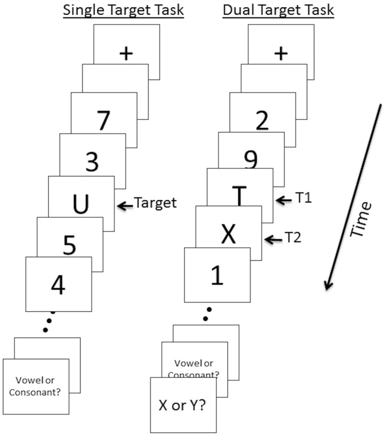
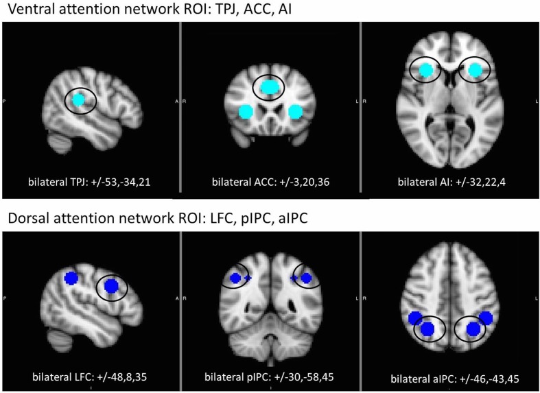
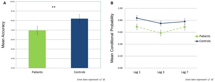
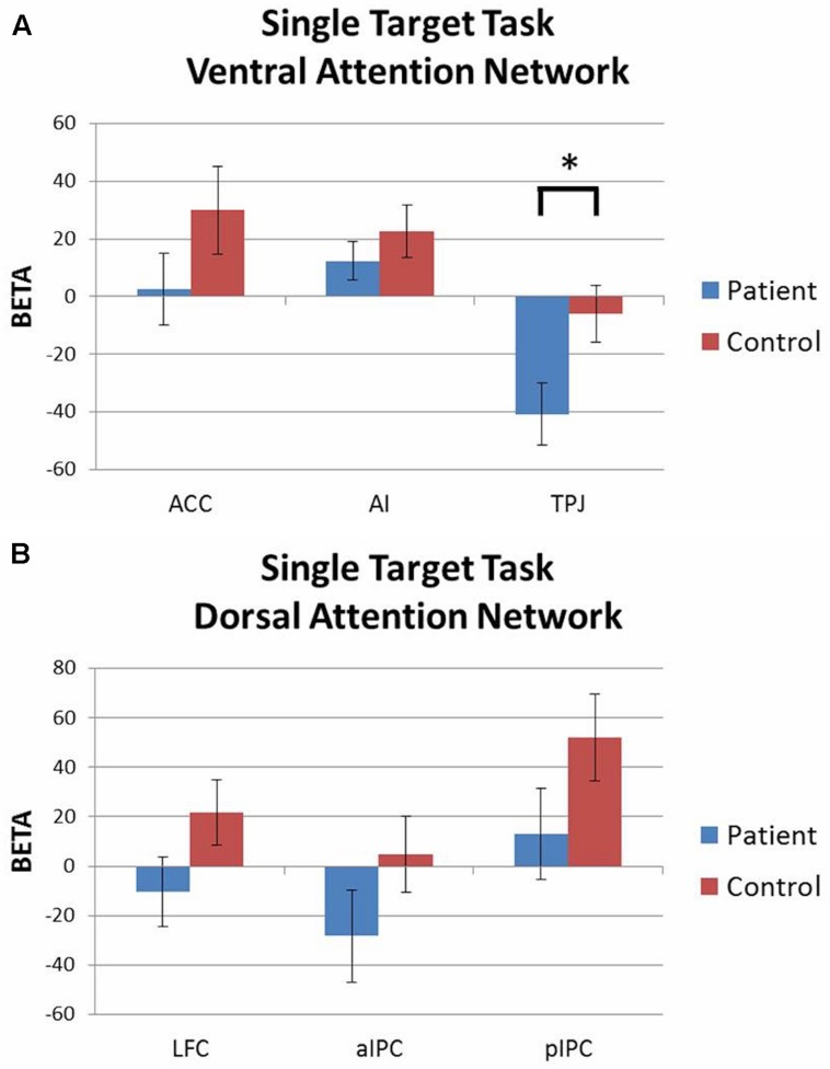
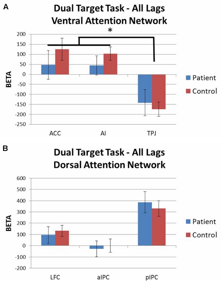
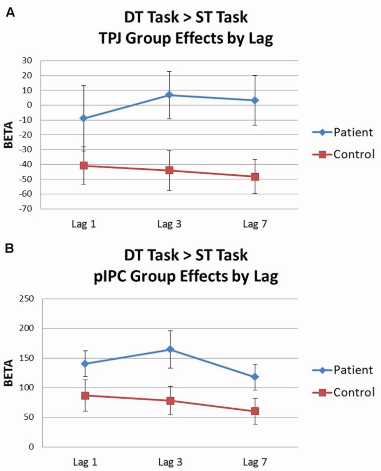
Early visual perception and attention are impaired in schizophrenia, and these deficits can be observed on target detection tasks. These tasks activate distinct ventral and dorsal brain networks which support stimulus-driven and goal-directed attention, respectively. We used single and dual target rapid serial visual presentation (RSVP) tasks during fMRI with an ROI approach to examine regions within these networks associated with target detection and the attentional blink (AB) in 21 schizophrenia outpatients and 25 healthy controls. In both tasks, letters were targets and numbers were distractors. For the dual target task, the second target (T2) was presented at three different lags after the first target (T1) (lag1 = 100 ms, lag3 = 300 ms, lag7 = 700ms). For both single and dual target tasks, patients identified fewer targets than controls. For the dual target task, both groups showed the expected AB effect with poorer performance at lag 3 than at lags 1 or 7, and there was no group by lag interaction. During the single target task, patients showed abnormally increased deactivation of the temporo-parietal junction (TPJ), a key region of the ventral network. When attention demands were increased during the dual target task, patients showed overactivation of the posterior intraparietal cortex, a key dorsal network region, along with failure to deactivate TPJ. Results suggest inefficient and faulty suppression of salience-oriented processing regions, resulting in increased sensitivity to stimuli in general, and difficulty distinguishing targets from non-targets.
Keywords: RSVP; attentional blink; fMRI; schizophrenia; visual attention.
Figures
Similar articles
-
Deficits and compensation: Attentional control cortical networks in schizophrenia.Neuroimage Clin. 2020;27:102348. doi: 10.1016/j.nicl.2020.102348. Epub 2020 Jul 20. Neuroimage Clin. 2020. PMID: 32736323 Free PMC article.
-
The attentional blink in schizophrenia: isolating the perception/attention interface.J Psychiatr Res. 2011 Oct;45(10):1346-51. doi: 10.1016/j.jpsychires.2011.04.002. Epub 2011 May 6. J Psychiatr Res. 2011. PMID: 21550051 Free PMC article.
-
A comparison between schizophrenia patients and healthy controls on the expression of attentional blink in a rapid serial visual presentation (RSVP) paradigm.Schizophr Bull. 2002;28(3):443-58. doi: 10.1093/oxfordjournals.schbul.a006952. Schizophr Bull. 2002. PMID: 12645676
-
Functional connectivity when detecting rare visual targets in schizophrenia.Psychiatry Res Neuroimaging. 2017 Mar 30;261:35-43. doi: 10.1016/j.pscychresns.2017.01.007. Epub 2017 Jan 17. Psychiatry Res Neuroimaging. 2017. PMID: 28126618 Free PMC article.
-
Behavioral cartography of visual functions in cat parietal cortex: areal and laminar dissociations.Prog Brain Res. 2001;134:265-84. doi: 10.1016/s0079-6123(01)34018-9. Prog Brain Res. 2001. PMID: 11702548 Review.
Cited by
-
High-definition transcranial direct current stimulation of the occipital cortices induces polarity dependent effects within the brain regions serving attentional reorientation.Hum Brain Mapp. 2022 Apr 15;43(6):1930-1940. doi: 10.1002/hbm.25764. Epub 2022 Jan 7. Hum Brain Mapp. 2022. PMID: 34997673 Free PMC article.
-
Deficits and compensation: Attentional control cortical networks in schizophrenia.Neuroimage Clin. 2020;27:102348. doi: 10.1016/j.nicl.2020.102348. Epub 2020 Jul 20. Neuroimage Clin. 2020. PMID: 32736323 Free PMC article.
-
Study of brain network alternations in non-lesional epilepsy patients by BOLD-fMRI.Front Neurosci. 2023 Jan 18;16:1031163. doi: 10.3389/fnins.2022.1031163. eCollection 2022. Front Neurosci. 2023. PMID: 36741055 Free PMC article.
-
Involvement of the dorsal and ventral attention networks in visual attention span.Hum Brain Mapp. 2022 Apr 15;43(6):1941-1954. doi: 10.1002/hbm.25765. Epub 2022 Jan 4. Hum Brain Mapp. 2022. PMID: 34984762 Free PMC article.
-
Switching between External and Internal Attention in Hippocampal Networks.J Neurosci. 2023 Sep 20;43(38):6538-6552. doi: 10.1523/JNEUROSCI.0029-23.2023. Epub 2023 Aug 22. J Neurosci. 2023. PMID: 37607818 Free PMC article.
References
-
- Cheung V., Chen E. Y. H., Chen R. Y. L., Woo M. F., Yee B. K. (2002). A comparison between schizophrenia patients and healthy controls on the expression of attentional blink in a rapid serial visual presentation (RSVP) paradigm. Schizophr. Bull. 28 443–458. 10.1093/oxfordjournals.schbul.a006952 - DOI - PubMed
Grants and funding
LinkOut - more resources
Full Text Sources
Other Literature Sources