Silver As Antibacterial toward Listeria monocytogenes
- PMID: 27014230
- PMCID: PMC4779933
- DOI: 10.3389/fmicb.2016.00307
Silver As Antibacterial toward Listeria monocytogenes
Abstract
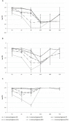
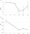
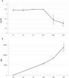
Listeria monocytogenes is a serious foodborne pathogen that can contaminate food during processing and can grow during food shelf-life. New types of safe and effective food contact materials embedding antimicrobial agents, like silver, can play an important role in the food industry. The present work aimed at evaluating the in vitro growth kinetics of different strains of L. monocytogenes in the presence of silver, both in its ionic and nano form. The antimicrobial effect was determined by assaying the number of culturable bacterial cells, which formed colonies after incubation in the presence of silver nanoparticles (AgNPs) or silver nitrate (AgNO3). Ionic release experiments were performed in parallel. A different reduction of bacterial viability between silver ionic and nano forms was observed, with a time delayed effect exerted by AgNPs. An association between antimicrobial activity and ions concentration was shown by both silver chemical forms, suggesting the major role of ions in the antimicrobial mode of action.
Keywords: cross-contamination; food preservation; food safety; foodborne pathogens; nanoparticles.
Figures


References
-
- Beigmohammadi F., Peighambardoust S. H., Hesari J., Azadmard-Damirchi S., Peighambardoust S. J., Khosrowshahi N. K. (2016). Antibacterial properties of LDPE nanocomposite films in packaging of UF cheese. LWT Food Sci. Technol. 65 106–111. 10.1016/j.lwt.2015.07.059 - DOI
-
- Berton V., Montesi F., Losasso C., Facco D. R., Toffan A., Terregino C. (2015). Study of the interaction between silver nanoparticles and Salmonella as revealed by transmission electron microscopy. J. Prob. Health 03 123 10.4172/2329-8901.1000123 - DOI
LinkOut - more resources
Full Text Sources
Other Literature Sources
Research Materials

