

Does Congenital Deafness Affect the Structural and Functional Architecture of Primary Visual Cortex?
- PMID: 27014392
- PMCID: PMC4787313
- DOI: 10.2174/1874440001610010001
Does Congenital Deafness Affect the Structural and Functional Architecture of Primary Visual Cortex?
Abstract
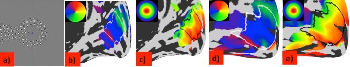
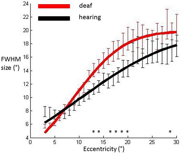
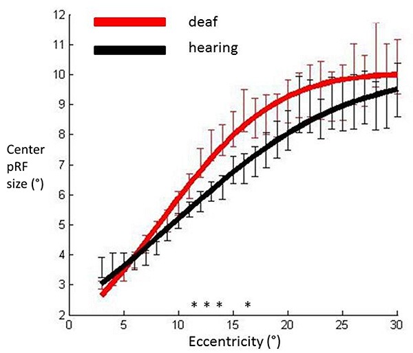
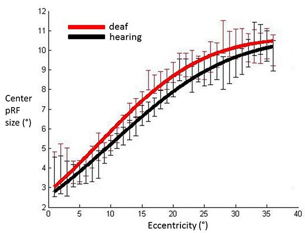
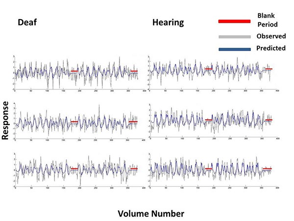
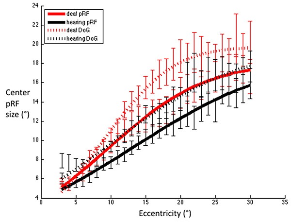
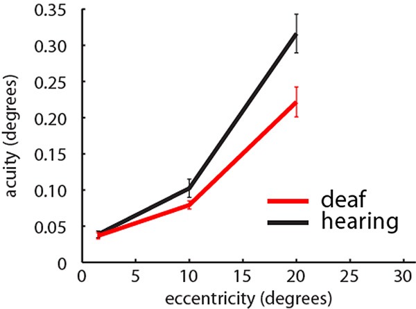
Deafness results in greater reliance on the remaining senses. It is unknown whether the cortical architecture of the intact senses is optimized to compensate for lost input. Here we performed widefield population receptive field (pRF) mapping of primary visual cortex (V1) with functional magnetic resonance imaging (fMRI) in hearing and congenitally deaf participants, all of whom had learnt sign language after the age of 10 years. We found larger pRFs encoding the peripheral visual field of deaf compared to hearing participants. This was likely driven by larger facilitatory center zones of the pRF profile concentrated in the near and far periphery in the deaf group. pRF density was comparable between groups, indicating pRFs overlapped more in the deaf group. This could suggest that a coarse coding strategy underlies enhanced peripheral visual skills in deaf people. Cortical thickness was also decreased in V1 in the deaf group. These findings suggest deafness causes structural and functional plasticity at the earliest stages of visual cortex.
Keywords: Deafness; functional magnetic resonance imaging (fMRI); peripheral visual field (PVF); primary visual cortex (V1).
Figures

References
-
- Wiesel T.N., Hubel D.H. Single cell responses of striate cortex of kittens deprived of vision in one eye. J. Neurophysiol. 1963;26:1003–1017. - PubMed
-
- Raemaekers M., Bergsma D.P., van Wezel R.J., van der Wildt G.J., van den Berg A.V. Effects of vision restoration training on early visual cortex in patients with cerebral blindness investigated with functional magnetic resonance imaging. J. Neurophysiol. 2011;105(2):872–882. doi: 10.1152/jn.00308.2010. - DOI - PubMed
Grants and funding
LinkOut - more resources
Full Text Sources
Other Literature Sources