. 2016 Jan;7(1-2):47-58.
doi: 10.18632/genesandcancer.94.

Expression profile of Wilms Tumor 1 (WT1) isoforms in undifferentiated and all-trans retinoic acid differentiated neuroblastoma cells

Affiliations

Expression profile of Wilms Tumor 1 (WT1) isoforms in undifferentiated and all-trans retinoic acid differentiated neuroblastoma cells

Grazia Maugeri et al. Genes Cancer. 2016 Jan.

Abstract

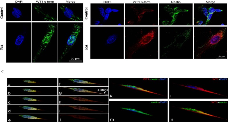
Wilms tumor 1 gene (WT1) is a tumor suppressor gene originally identified in nephroblastoma. It is also expressed in neuroblastoma which represents the most aggressive extracranial pediatric tumor. Many evidences have shown that neuroblastoma may undergo maturation, by transforming itself in a more differentiated tumors such as ganglioneuroblastoma and ganglioneuroma, or progressing into a highly aggressive metastatic malignancy. To date, 13 WT1 mRNA alternative splice variants have been identified. However, most of the studies have focused their attention only on isoform of ∼49 kDa. In the present study, it has been investigated the expression pattern of WT1 isoforms in an in vitro model of neuroblastoma consisting in undifferentiated or all-trans retinoic acid (RA) differentiated cells. These latter representing the less malignant phenotype of this tumor. Results have demonstrated that WT1.1-WT1.5, WT1.6-WT1.9, WT1.10 WT1.11-WT1.12 and WT1.13 isoforms are expressed in both groups of cells, but their levels are significantly increased after RA treatment. These data have also been confirmed by immunofluorescence analysis. Moreover, the inhibition of PI3K/Akt and MAPK/ERK, that represent two signalling pathway specifically involved in NB differentiation, induces an overexpression of WT1 isoforms. These data suggest that WT1 isoforms might be involved in differentiation of neuroblastic into mature ganglion cells.

Keywords: PI3K/Akt and MAPK/ERK signalling pathway; WT1 isoforms; Wilm's tumor 1 gene; alternative splice variants; neuroblastoma; tumor suppressor gene.


Conflict of interest statement

CONFLICTS OF INTERESTS

The authors declare that they have no competing interests.

Figures

Figure 1
Figure 1. Alignment of WT1 isoforms
Multiple alignment of WT1 isoforms showing the matching regions to canonical sequence WT1.6.
Figure 2
Figure 2. Expression profile of WT1 isoforms in undifferentiated and RA differentiated neuroblastoma cells
Representative immunoblot of WT1 isoforms expression on lysate from SH-SY5Y cells cultured in growth medium (Control) or added with 10 μM all-trans retinoic acid (RA), detected by WT1 C-terminal (A) and WT1 N-terminal antibody (C). Blot shows the results of three independent experiments. The boxes frame signals detected from each isoform with relative ID Code, as listed in Table 1. (B-D) Relative density of each band was quantified using ImageJ software. Each signal was normalized on correspondent β-tubulin signal. Data are expressed as mean ± S.E.M. ***p<0,001 vs Control, as determined by unpaired two-tailed Student t-test.
Figure 3
Figure 3. Representative 2D gel immunoblots of WT1 protein isoforms expressed in undifferentiated and RA differentiated neuroblastoma cells
The images are representative of two-dimensional electrophoresis blots showing WT1 signals detected on lysate from SH-SY5Y cells cultured in growth medium (Control) or added with 10 μM all-trans retinoic acid (RA), detected by WT1 C-terminal and WT1 N-terminal antibody. The blots are representative of three independent experiments.
Figure 4
Figure 4. Nestin expression in undifferentiated and RA differentiated neuroblastoma cells
(A) Representative immunoblots of nestin expression on lysate from SH-SY5Y cells cultured in growth medium (Control) or added with 10 μM all-trans retinoic acid (RA). Blot shows the results of three independent experiments. (B) Relative density of each band was quantified using ImageJ software. Each signal was normalized on correspondent β-tubulin signal. Data are expressed as mean ± S.E.M. ***p<0,001 vs Control as determined by unpaired two-tailed Student t-test.
Figure 5
Figure 5. Phosphorylation of AKT and ERK1/2 in undifferentiated and RA differentiated neuroblastoma cells
(A) Phosphorylation of Ser473Akt in SH-SY5Y cells cultured in growth medium (Control) or added with 10 μM all-trans retinoic acid (RA), or with 10 μM Wortmannin (Wortmannin), or with 10 μM all-trans retinoic acid and 10 μM Wortmannin (RA+Wortmannin), as determined by Western blots. Relative density of each band was quantified using ImageJ software. Each signal of phosphorylated protein was normalized to total protein expression. Data are expressed as mean ± S.E.M. ***p<0,001 vs Control; ###p<0,001 vs RA as determined by one-way ANOVA followed by the Tukey post hoc test. (B) Phosphorylation of Erk1/2 in SH-SY5Y cells cultured in growth medium (Control) or added with 10 μM all-trans retinoic acid (RA), or added with 50 μM PD98059, or with 10 μM all-trans retinoic acid and 50 μM PD98059 (RA+PD98059), as determined by Western blots. Relative density of each band was quantified using ImageJ software. Each signal of phosphorylated protein was normalized to total protein expression. Data are expressed as mean ± S.E.M. ***p<0,001 vs Control; ###p<0,001 vs RA as determined by one-way ANOVA followed by the Tukey post hoc test.
Figure 6
Figure 6. Expression profile of WT1 isoforms after inhibition of the phosphoinositide 3-kinase (PI3K)/Akt signaling pathway or of the mammalian mitogen activated protein kinase/Erk kinase (MAPPK or MEK) family
Representative immunoblot of WT1 isoforms expression, detected by WT1 C-terminal (A) and WT1 N-terminal (B) antibody, on lysate from SH-SY5Y cells cultured in growth medium (Control), or added with 10 μM all-trans retinoic acid (RA), or with 10 μM Wortmannin (Wortmannin), or with 50 μM PD98059 (PD98059), or with 10 μM all-trans retinoic acid and 10 μM Wortmannin (RA+Wortmannin), or with 10 μM all-trans retinoic acid and 50 μM PD98059 (RA+PD98059). (C-G) Relative density of each band was quantified using ImageJ software. Each signal was normalized on correspondent β-tubulin signal. Data are expressed as mean ± S.E.M. ***p<0,001 vs Control; ###p<0,001 vs RA as determined by one-way ANOVA followed by the Tukey post hoc test.
Figure 7
Figure 7. Immunolocalization of WT1 isoforms and Nestin in undifferentiated and RA differentiated neuroblastoma cells
(A) Representative photomicrographs show total WT1 isoforms detected by WT1 C-terminal antibody (green) in SH-SY5Y cells. Nuclei were stained with DAPI (blue). (B) Representative photomicrographs show immunolocalization of total WT1 isoforms detected by WT1 N-terminal antibody (red) and Nestin protein (green) in SH-SY5Y cells. Nuclei were stained with DAPI (blue). (C) Immunolocalization of total WT1 isoforms detected by WT1 N-terminal antibody (red) and Nestin protein (green). a to j: serial z-sections (steps of 0.5 μm) obtained by confocal laser scanning microscope. k to n: image along the z-plane indicated in the panel g. Cells were cultured in growth medium (Control) or added with 10 μM all-trans retinoic acid (RA). Photomicrographs are representative results of fields taken randomly from slide and scanned by confocal laser scanning microscopy (CLSM; Zeiss LSM700).

References

    1. Call KM, Glaser T, Ito CY, Buckler AJ, Pelletier J, Haber DA, Rose EA, Kral A, Yeger H, Lewis WH, Jones C, Housman DE. Isolation and characterization of a zinc finger polypeptide gene at the human chromosome 11 Wilms' tumor locus. Cell. 1990;60:509–520. - PubMed
    1. Gessler M, Poustka A, Cavenee W, Neve RL, Orkin SH, Bruns GAP. Homozygous deletion in Wilms' tumors of a zinc-finger gene identified by chromosome jumping. Nature. 1990;343:774–8. - PubMed
    1. Scharnhorst V, van der Eb AJ, Jochemsen AG. WT1 proteins: functions in growth and differentiation. Gene. 2001;273:141–61. - PubMed
    1. Menke AL, van der Eb AJ, Jochemsen AG. The Wilms' tumor 1 gene: oncogene or tumor suppressor gene? Int Rev Cytol. 1998;181:151–212. - PubMed
    1. Yang L, Han Y, Suarez Saiz F, Minden MD. A tumor suppressor and oncogene: the WT1 story. Leukemia. 2007;21:868–876. - PubMed

LinkOut - more resources