

Update on three-dimensional image reconstruction for preoperative simulation in thoracic surgery
- PMID: 27014477
- PMCID: PMC4783726
- DOI: 10.3978/j.issn.2072-1439.2016.02.39
Update on three-dimensional image reconstruction for preoperative simulation in thoracic surgery
Abstract
Background: Three-dimensional computed tomography (3D-CT) technologies have been developed and refined over time. Recently, high-speed and high-quality 3D-CT technologies have also been introduced to the field of thoracic surgery. The purpose of this manuscript is to demonstrate several examples of these 3D-CT technologies in various scenarios in thoracic surgery.
Methods: A newly-developed high-speed and high-quality 3D image analysis software system was used in Kyoto University Hospital. Simulation and/or navigation were performed using this 3D-CT technology in various thoracic surgeries.
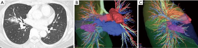
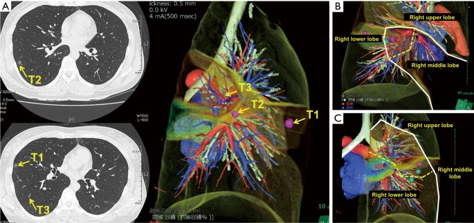
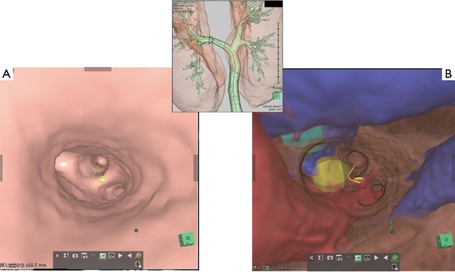
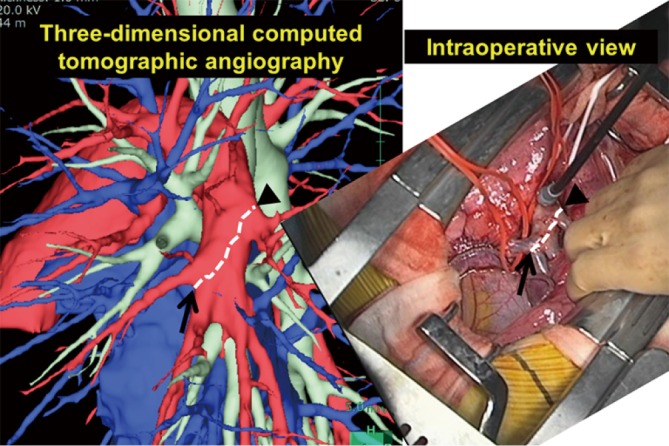
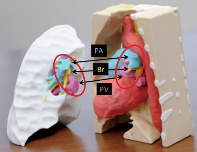
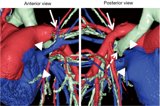
Results: Preoperative 3D-CT simulation was performed in most patients undergoing video-assisted thoracoscopic surgery (VATS). Anatomical variation was frequently detected preoperatively, which was useful in performing VATS procedures when using only a monitor for vision. In sublobar resection, 3D-CT simulation was more helpful. In small lung lesions, which were supposedly neither visible nor palpable, preoperative marking of the lesions was performed using 3D-CT simulation, and wedge resection or segmentectomy was successfully performed with confidence. This technique also enabled virtual-reality endobronchial ultrasonography (EBUS), which made the procedure more safe and reliable. Furthermore, in living-donor lobar lung transplantation (LDLLT), surgical procedures for donor lobectomy were simulated preoperatively by 3D-CT angiography, which also affected surgical procedures for recipient surgery. New surgical techniques such as right and left inverted LDLLT were also established using 3D models created with this technique.
Conclusions: After the introduction of 3D-CT technology to the field of thoracic surgery, preoperative simulation has been developed for various thoracic procedures. In the near future, this technique will become more common in thoracic surgery, and frequent use by thoracic surgeons will be seen in worldwide daily practice.
Keywords: Endobronchial ultrasonography (EBUS); living-donor lobar lung transplantation (LDLLT); thoracic surgery; three-dimensional computed tomography (3D-CT); video-assisted thoracoscopic surgery (VATS).
Conflict of interest statement
Figures
References
-
- Yamanaka J, Saito S, Iimuro Y, et al. The impact of 3-D virtual hepatectomy simulation in living-donor liver transplantation. J Hepatobiliary Pancreat Surg 2006;13:363-9. - PubMed
-
- Mochizuki K, Takatsuki M, Soyama A, et al. The usefulness of a high-speed 3D-image analysis system in pediatric living donor liver transplantation. Ann Transplant 2012;17:31-4. - PubMed
-
- Ikeda N, Yoshimura A, Hagiwara M, et al. Three dimensional computed tomography lung modeling is useful in simulation and navigation of lung cancer surgery. Ann Thorac Cardiovasc Surg 2013;19:1-5. - PubMed
-
- Sato M, Omasa M, Chen F, et al. Use of virtual assisted lung mapping (VAL-MAP), a bronchoscopic multispot dye-marking technique using virtual images, for precise navigation of thoracoscopic sublobar lung resection. J Thorac Cardiovasc Surg 2014;147:1813-9. - PubMed
-
- Herth FJ, Annema JT, Eberhardt R, et al. Endobronchial ultrasound with transbronchial needle aspiration for restaging the mediastinum in lung cancer. J Clin Oncol 2008;26:3346-50. - PubMed
LinkOut - more resources
Full Text Sources