Review
. 2016 Mar 9:6:28.
doi: 10.3389/fcimb.2016.00028. eCollection 2016.

Implications of Spatiotemporal Regulation of Shigella flexneri Type Three Secretion Activity on Effector Functions: Think Globally, Act Locally

Affiliations
Review

Implications of Spatiotemporal Regulation of Shigella flexneri Type Three Secretion Activity on Effector Functions: Think Globally, Act Locally

F-X Campbell-Valois et al. Front Cell Infect Microbiol. .

Abstract

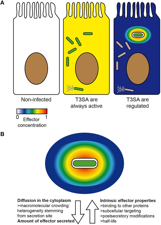
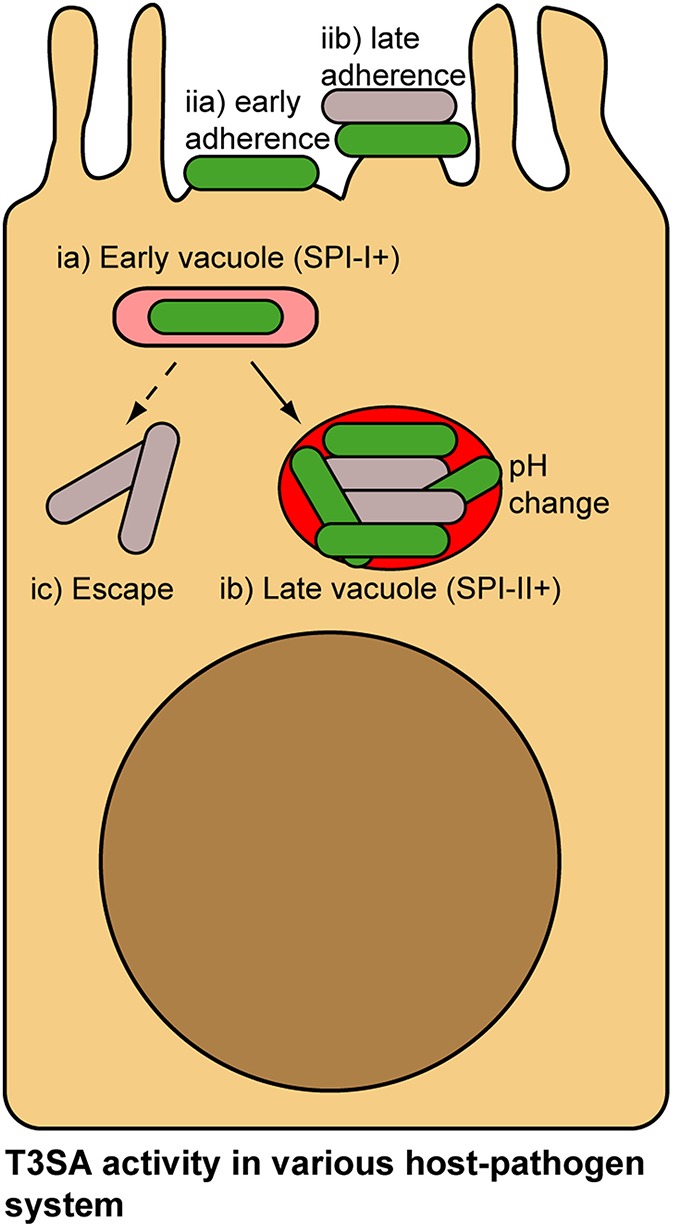
Shigella spp. are Gram-negative bacterial pathogens that infect human colonic epithelia and cause bacterial dysentery. These bacteria express multiple copies of a syringe-like protein complex, the Type Three Secretion apparatus (T3SA), which is instrumental in the etiology of the disease. The T3SA triggers the plasma membrane (PM) engulfment of the bacteria by host cells during the initial entry process. It then enables bacteria to escape the resulting phagocytic-like vacuole. Freed bacteria form actin comets to move in the cytoplasm, which provokes bacterial collision with the inner leaflet of the PM. This phenomenon culminates in T3SA-dependent secondary uptake and vacuolar rupture in neighboring cells in a process akin to what is observed during entry and named cell-to-cell spread. The activity of the T3SA of Shigella flexneri was recently demonstrated to display an on/off regulation during the infection. While the T3SA is active when bacteria are in contact with PM-derived compartments, it switches to an inactive state when bacteria are released within the cytosol. These observations indicate that effector proteins transiting through the T3SA are therefore translocated in a highly time and space constrained fashion, likely impacting on their cellular distribution. Herein, we present what is currently known about the composition, the assembly and the regulation of the T3SA activity and discuss the consequences of the on/off regulation of T3SA on Shigella effector properties and functions during the infection. Specific examples that will be developed include the role of effectors IcsB and VirA in the escape from LC3/ATG8-positive vacuoles formed during cell-to-cell spread and of IpaJ protease activity against N-miristoylated proteins. The conservation of a similar regulation of T3SA activity in other pathogens such as Salmonella or Enteropathogenic Escherichia coli will also be briefly discussed.

Keywords: Shigella; effectors; enteropathogens; host-pathogen interactions; signal transduction; type three secretion apparatus; type three secretion system.


Figures

Figure 1
Figure 1
Scheme of the type three secretion apparatus of Shigella flexneri. Basic scheme of Shigella flexneri T3SA at permissive temperatures (e.g., 37°C) in the inactive (A) and active states (B). The tip complex is composed of IpaD (grey circles) and IpaB (white rectangles) adopting a closed conformation and an open conformation in the inactive and active states, respectively. Activation of secretion leads to MxiE-IpgC-dependent expression of second wave effectors. The dashed arrow indicates the route followed by translocators and effectors during secretion. They travel through a conduit located at the center of the T3SA that comprises successively the sorting platform, the inner membrane ring, rod protein (not visible on this scheme), the outer membrane ring, the needle, and translocon. Numbers in parenthesis in the right panel indicate the secretion order of translocators, first wave and second wave effectors. Labelings of bacterial cytoplasmic complex components are indicated from left to right. IM, inner membrane; OM, outer membrane; HPM, host plasma membrane.
Figure 2
Figure 2
Shigella infectious cycle: interaction of T3SA with membrane compartments is key for the regulation of its activity. The “invade and evade” infectious strategy of S. flexneri can be broken down in two phases: (1) entry, characterized by residence of bacteria in vacuoles derived from the PM (1a,b), which are ultimately ruptured (1c); (2) cytoplasmic residence, where most replication events occur (2a) and motility through actin comet formation is possible (2b). After bacteria have reached the cytoplasm, they are in position to iterate this cycle and progressively invade neighboring cells, before evading once again the secondary vacuole. This process is characterized by the formation of protrusions (3a) and vacuoles (3b) composed of a double membrane derived from the PM in which bacteria reside until their lysis (3c), and escape in the cytoplasm (2*). It was demonstrated that secreting bacteria (green) were systematically associated with entry and cell-to-cell spread vacuoles and protrusions derived from the PM, while cytoplasmic bacteria were not actively secreting (gray) (A). Magnification of the inner and outer leaflet of the PM. Density of cholesterol (yellow rectangles) and overall phospholipids composition (pink vs. blue) is variable in both leaflets. Therefore, bacteria are not facing the same biochemical cues when they are performing entry vs. cell-to-cell spread. As mentioned in panel (A), bacteria also face four membranes during cell-to-cell spread instead of two during entry (B). Proposed mechanisms of inactivation of T3SA in intracellular Shigella. Grey circles and white rectangles represent secreted tip complex proteins, which are incapable of blocking T3SA conduit. H1 and H2 represent alternative hypotheses for inactivation of T3SA in the cytoplasm of host cells, as described in the text. H1: replenishment of functional tip complex; H2: disassembly of T3SA before vacuole escape and replenishment with inactive T3SA in the host cytoplasm (C).
Figure 3
Figure 3
Factors impacting on the concentration and distribution of effectors inside host cells. Theoretical concentration and distribution of bacterial effectors in an uninfected cell (left) vs. in infected cells in the case scenario where all intracellular bacteria (center) or only those in vacuoles are secreting (right) (A). An actively secreting bacterium located in a vacuole is represented with its theoretical gradient of effectors. Arrows oriented away and toward the bacteria represent respectively factors favoring or disfavoring rapid and homogenous diffusion of effectors inside host cells (B). Secreting bacteria, green; non-secreting bacteria, gray. Color scale represents concentration of effectors from 0 (min) to 1 (max) (arbitrary unit).
Figure 4
Figure 4
Local vs. distant action of secreted effectors. Factors influencing the balance between local and distant action of effectors from their secretion site inside host cells (A). Examples of local and distant action of effectors inside host cells. OspF is acting in the nucleus (distant action) to block the inflammatory response, while IcsB and VirA were recently proposed to act directly on the cell-to-cell spread vacuole (local action) to favor bacterial escape in the cytoplasm. Question marks indicate the possibility that OspF and IcsB/VirA could also have local and distant functions, respectively (B).
Figure 5
Figure 5
Alternative lifestyles of pathogenic bacteria associated with host cells and potential mechanisms of regulation of their T3SA. Salmonella invade epithelial cells using Salmonella Pathogenicity Island-1 (SPI-1) T3SA (ia). pH change in the vacuole and concomitant sensing of cytosolic pH induce activation of Salmonella Pathogenicity Island-2 (SPI-2) T3SA in Salmonella (ib). As bacteria accumulate in vacuole, they are not in contact with the membrane, which would probably inactivate SPI-1 and SPI-2 T3SA (ib). Occasionally Wt Salmonella escape their vacuole and access the cytoplasm. Loss of contact with vacuolar membrane in this case could also potentially lead to T3SA inactivation (ic). EPEC adhesion to the PM of epithelial cells proceeds in multiple stages (e.g., bundling forming pili-, T3SS/Tir- and EspA-dependent, etc.), culminating in the formation of actin-rich pedestals structure at bacterial adhesion point. Throughout this adhesion process, activity of the T3SA could be modulated (iia,b). In addition, within microcolonies some bacteria will occasionally loose contact with the PM, which similarly to the previous example could inactivate T3SA. Secreting bacteria, green; non-secreting bacteria, gray.

References

    1. Ambrosi C., Pompili M., Scribano D., Limongi D., Petrucca A., Cannavacciuolo S., et al. . (2015). The Shigella flexneri OspB effector: an early immunomodulator. Int. J. Med. Microbiol. 305, 75–84. 10.1016/j.ijmm.2014.11.004 - DOI - PubMed
    1. Arbibe L., Kim D. W., Batsche E., Pedron T., Mateescu B., Muchardt C., et al. . (2007). An injected bacterial effector targets chromatin access for transcription factor NF-kappaB to alter transcription of host genes involved in immune responses. Nat. Immunol. 8, 47–56. 10.1038/ni1423 - DOI - PubMed
    1. Arena E. T., Campbell-Valois F.-X., Tinevez J.-Y., Nigro G., Sachse M., Moya-Nilges M., et al. . (2015). Bioimage analysis of Shigella infection reveals targeting of colonic crypts. Proc. Natl. Acad. Sci. U.S.A. 112, E3282–E3290. 10.1073/pnas.1509091112 - DOI - PMC - PubMed
    1. Ashida H., Kim M., Sasakawa C. (2014). Exploitation of the host ubiquitin system by human bacterial pathogens. Nat. Rev. Microbiol. 12, 399–413. 10.1038/nrmicro3259 - DOI - PubMed
    1. Ashida H., Kim M., Schmidt-Supprian M., Ma A., Ogawa M., Sasakawa C. (2010). A bacterial E3 ubiquitin ligase IpaH9.8 targets NEMO/IKKgamma to dampen the host NF-kappaB-mediated inflammatory response. Nat. Cell Biol. 12, 66–73; sup pp 1–9. 10.1038/ncb2006 - DOI - PMC - PubMed

Publication types

MeSH terms

Substances